NEONATOLOGY ON THE WEB
 |
Asphyxia Neonatorum: Causation and
Treatment.
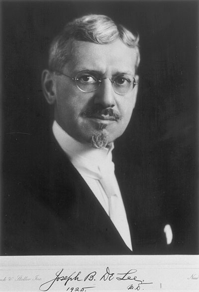
By Jos. B. De Lee, M.D., Chicago.
Professor of Obstetrics, Northwestern University Medical School;
Obstetrician to Mercy Hospital and to the Chicago Lying-in Hospital.
Published in Medicine (Detroit) 3:643-660, 1897.
Read at the forty-seventh annual meeting of the Illinois State
Medical Society, held in East St. Louis, May 18, 1897.
|
This is a subject of daily interest to the general practitioner,
of special interest to the obstetrician; it is acquiring importance
in the eyes of the alienist and pediatrician, and to the general
hygienist its consideration has become of great moment.
The highest mortality that befalls the human race in one day
occurs on the day of birth. Schultze [1]
estimates that five per cent. of children are still-born, dying
during labor, and one and a half per cent. die shortly after birth,
the result of the trauma of labor. Brothers [2]
said that in New York City, in the four years 1889 to 1892, over
16,000 children were born dead or died immediately after birth. I
believe these figures are not too large. It is said that the most
important period of the life of a human being is the time spent in
utero. The most trying ordeal a human being sustains is the
ordeal of birth.
The fetus receiving food and oxygen from the mother while in the
uterus is suddenly made dependent on outer conditions for these
necessities. While during the short period of labor the absence of
food will have no influence, the withdrawal of oxygen becomes of
immediate and serious moment to the fetus.
Many children die in the first days because too little food is
taken or it is not assimilated, but as many more die during labor
because of the interruption of the respiratory function -- die of
asphyxia.
It is a fact that can be clinically attested daily that during the
uterine contraction -- the labor pain -- the heart-tones of the fetus
sink in frequency and force. During a long pain the beats may become
very few or even inaudible. While this in part is due to the muscular
sound of the uterus interfering with the transmission of the fetal
heart-beats, one can notice as the pain passes off a rapid return to
the normal frequency and force. After the rupture of the bag of
waters this effect of the pain is much intensified. This action of
the uterine contraction on the heart has been long known and is
constant. The circulation of the blood in the placenta is interfered
with as a result of the compression of the uterine contents, the
interchange of gases between the fetal and maternal blood is stopped,
the absence of oxygen in the fetal blood irritates the vagus -- the
pneumogastric nerve -- and the heart is therefore slowed. Experiments
on dogs show this; by interrupting the respiration of the dog put in
a state of apnoea by artificial respiration with the bellows, the
heart is slowed.
Toward the end of the second stage the heart-tones are much
diminished by the pains, and during the passage of the head through
the vulva they may be sixty per minute. This is due to the great
compression of the placenta, especially marked after the rupture of
the bag of waters; to the beginning separation of the placenta, which
normally occurs at the end of the second stage of labor; and to a
certain extent to compression of the brain. We have here the common
cause of asphyxia. Too frequent and powerful labor pains, prolonged,
with short intervals of relaxation, are very dangerous to fetal life.
Very striking in this regard is the bad effect of ergot. This drug
lengthens the pains, lessens the intervals, and increases the force
of the uterine contraction; it sometimes even produces tetanus uteri,
under which fetal life is quickly extinguished. The same dangers
menace the fetus when from too frequent manipulation or attempts at
delivery the pains assume a tetanic character.
Since in cases of suffocation of the pregnant woman the fetus dies
first because the mother abstracts oxygen from the fetal circulation,
this and other conditions in the mother -- as anemia, acute and
chronic, heart and lung disease, asphyxiation by gas and so forth --
are dangerous for the child. It is this that makes the prognosis bad
for children delivered by Caesarian section from women who die
slowly. After the sudden death of the mother the fetus may live
fifteen minutes, and if promptly extracted may recover. Only six per
cent. of the hitherto reported cases have lived.
Premature separation of the placenta normally implanted may happen
during pregnancy, and then is almost always fatal to the child; or it
may occur near the end of the second stage of labor, when the child
may be born alive. The death of the second twin in utero is
often caused by too early separation of the common placenta. Besides
the signs of asphyxiation shown by the fetus, external hemorrhage in
the interval between the births gives warning of the danger.
Separation of the placenta when it is praevia, or compression of the
forelying placenta by the breech, or colpeurynter used to stop
hemorrhage, causes asphyxia. When the placenta is torn by
manipulation anemia is added to the asphyxia, and these are further
aggravated by the anemia of the mother -- all of which explain the
high fetal mortality in placenta praevia.
All those conditions which cause a compression of the cord will
obviously cause asphyxia, and in degree and rapidity according to the
degree and suddenness of the compression. Prolapsed cord comes first
in order of frequency. When the cord is coiled around the neck or any
part of the child the danger is great from compression or tension
[3] of the cord, or compression of the
vessels of the neck, and in the latter instance the tip of the
forceps may directly compress the cord. This point deserves special
emphasis and sounds the warning to carefully auscultate the
heart-tones during forceps delivery. In breech cases the funis may be
compressed between the trunk and the passages, but here compression
of the placenta by the relatively hard head has something to do with
the asphyxia.
Finally compression of the brain brings about asphyxia. During the
passage of the head through the vulva the slowing of the fetal heart
is observed and is due partly to this cause. When the head is being
forced into a contracted pelvis by strong pains, or when the head is
compressed in the blades of the forceps, even in easy deliveries a
marked slowing of the pulse can be noted. This is due to irritation
of the pneumogastric [vagus nerve]; as a result of the slowed
circulation the fetal blood becomes venous and a state of asphyxia is
inaugurated. In some cases of brain compression the acting cause is
so slow that the fetus dies without making any attempt at respiration
in the uterus. A fracture of the skull or an intracranial hemorrhage
brings the same result; but should the injury involve the medulla the
effect is worse, usually soon fatal. It is remarkable how in these
cases, while the respiratory center is paralyzed, the heart will
continue beating as long as oxygen is supplied by artificial
respiration.
While a fetus in the state of apnoea does not respond with
respiratory efforts to external stimuli, I have felt these motions on
introducing the hand to do a version, and have seen respiratory
motions follow traction on the foot in footling cases. Anesthetics
have been said to cause asphyxia, but extended experience with them
in both obstetric and surgical degrees has not demonstrated this.
The Treatment. -- Paramount in importance is the
recognition of asphyxia beginning while the child is still in
utero, and fortunately this is almost always possible. The cases
are very few where accurate information regarding the condition of
the child cannot be obtained. One of the first signs of fetal danger
is the decrease in the number of the heart-beats. A steady slowing to
one hundred per minute, and certainly if below this, points to
impending fetal danger. In some cases there is an increase in the
number to 160 or more. This usually comes on after the diminution has
been pronounced and betokens impending death from paralysis of the
vagus. It may be primary, however, and then should be accorded the
same significance as slowed heart's action. Irregularity of the
heart-tones occurring in the intervals between pains is also of
significance, and impurity of the first sound of the fetal heart
carries important information to the experienced ear.
The second diagnostic sign is the passage of meconium. In breech
cases this is of little value unless the breech is high up, and in
the other presentations the sign should be accorded significance only
when the meconium is fresh. It may easily happen that a transient
asphyxia has caused the fetus to lose its meconium, which may have
occurred weeks before. Liquor amnii stained with old meconium is
olive green in color, appears more watery, and the meconium is
thoroughly mixed with it. Fresh meconium is dark green, is lumpy, and
is not thoroughly mixed with the liquor amnii. Healthy fetuses have
passed meconium during labor. I once saw meconium come from the mouth
and nose of a child as the face escaped over the perineum. Quinine is
said to cause its discharge into the liquor amnii. Where the meconium
is passed as a result of asphyxia it is brought about, not by
paralysis of the sphincter ani, but by a hyperperistalsis caused by
the deoxygenated condition of the blood, and is analogous to the
involuntary bowel-movements so often observed in death by drowning,
strangling, etc.
In obstructed labors the passage of meconium may long precede the
slowing of the pulse, and thus is a valuable sign of beginning
danger. Until the discovery of the heart-tones this was the only
means possessed by the obstetrician to determine the condition of the
child.
Another sign, but one that shows that the asphyxia has already
become serious, is feeling, seeing, or hearing respiratory movements
made by the child. Where the breech is delivered one often sees the
gasps made by the child. After podalic version the foot is sometimes
seen to move with the respiratory action. In one case the effect of
fetal hiccough was thus observed. [4] During
a forceps delivery I once felt and saw the shoulders above the pubis
move in inspiration and expiration. One of the early signs of
asphyxia may be undue violent movements on the part of the child,
felt by the mother and attendant. Ordinarily the child is quiescent
during labor.
As everywhere else in medicine, but particularly in obstetrics,
prevention plays the most important rôle, and by it one can
accomplish much more than by treatment. One should recognize the
causes of asphyxia and avoid them. The bag of waters should not be
ruptured unnecessarily. While it is in the main true that the fetus
is in no danger of asphyxia in an intact sac, I have seen cases where
the heart-tones gave warning of fetal danger before the escape of the
waters. Upon rupturing the membranes meconium escaped, and the fetus
was delivered asphyxiated. Still these cases are few.
Ergot should not be given till after the placenta has been
expelled. In spite of the many pages that have been written against
the use of ergot during labor, it is still used, particularly by
midwives, to the destruction of many women and children. No attempt
should be made to hasten normal labor. The necessary manipulations in
many ways jeopardize the child. Particularly in breech and footling
cases should premature attempts at delivery be avoided. The
extraction begun before the cervix is fully dilated is usually
complicated by the stripping of the arms above the head, and the
delay caused in bringing them down is likely to be fatal.
[5]
Watch the heart-tones throughout the labor, especially toward the
end of the second stage and in all delayed labors. In operative
deliveries be ready to treat asphyxia neonatorum -- that is, have hot
water, hot towels, a bath, and the necessary instruments near at
hand.
When you have diagnosed asphyxia beginning in utero the
child must be delivered as rapidly as possible consistent with the
safety of the mother. Should the delivery be difficult the
heart-tones should be watched carefully, and should the fetal heart
cease beating before the delivery is accomplished embryotomy is to be
performed.
When a child is born apparently dead it is of instant importance
to determine whether the asphyxia be mild or severe. In mild asphyxia
-- asphyxia livida -- the child is rigid, blue, with turgid lips,
slight exophthalmos, and makes a few attempts at respiration, but the
mucus in the passages prevents air from entering the alveoli. A
finger in the throat provokes a reaction, and the heart beats
strongly. In severe asphyxia -- asphyxia pallida -- the child is
limp, the extremities hang down, the surface is pale except the lips,
the jaw falls or is jerked by an occasional gasp, the throat does not
react to the finger, the heart beats faintly or is only perceptible
to the ear. These are not different forms of the asphyxia, but
different degrees.
It is true that anemia of the fetus and compression or hemorrhage
into the brain present symptoms similar to those of asphyxia pallida,
but the treatment is the same since these conditions can usually not
be surely diagnosed. I have seen a case of morphine poisoning in a
child born of an eclamptic mother to whom large doses of the narcotic
had been given.
There are three grand principles governing the treatment of
asphyxia neonatorum: first, maintain the body heat; second, free the
air passages from obstructions; third, stimulate respiration, or
supply air to the lungs for oxygenation of the blood.
The importance of keeping the baby warm is not generally
appreciated. The infant is wet, exposure is often prolonged,
evaporation is rapid, the body temperature sinks rapidly. This is
very depressing; indeed, it has occasionally happened that an infant
wrapped up and put away as dead has recovered by the influence of
warmth alone. Therefore the baby should be wrapped in warm towels or
put in a warm full bath.
At the same time the air passages must be cleared. It is fruitless
to attempt to resuscitate a baby when the trachea, bronchi, and
sometimes the alveoli are full of amniotic fluid, meconium, blood, or
vaginal secretions. These must be removed before any attempt is made
to bring air into the lungs; otherwise the foreign substances would
be forced still farther down and give rise to atelectasis, pneumonia,
and sepsis.
In the mildest cases it is sufficient to wipe the throat with the
little finger covered with the corner of a soft towel while the child
is held up by the ankles. This also excites cough, and the mucus is
expelled. Attention is to be called to the statement of a recent
German observer [6] that there forms
normally in the glottis some mucus which is expelled after the birth
of the child, but which when sucked into the trachea may suffocate
it. In a forensic light this acquires some importance.
In the more severe cases -- asphyxia livida -- the deeper passages
must be cleared, and this is best done by means of a soft woven
catheter open at the end, No. 14 French. This little instrument ought
to be found in the satchel of every obstetrical attendant. With a
little practise its use becomes easy. The index finger of the left
hand pulls the epiglottis forward and comes to touch the arytenoid
cartilages. The catheter is then passed along the finger, the end is
pulled against the posterior surface of the epiglottis with the
finger, and is then pushed into the trachea with a gentle twisting
motion of the right hand. A slight suck draws the contents of the
trachea into the tube, it is withdrawn, and the mucus blown out on a
towel. This is repeated until the passages are clear.
In asphyxia livida this is almost always sufficient, the
irritation of the throat eliciting respirations. If these are slow in
coming a brisk rubbing of the back with a warm towel or in a hot bath
will stimulate them. Slapping on the back, cold water on the chest,
the cold plunge-bath, are almost never necessary, and the latter two
are distinctly harmful.
In the severest cases -- asphyxia pallida -- the diagnostic
criterion of which is the absence of reaction in the throat, this
simple treatment does not suffice. One must waste no time with trying
the skin reflexes, but immediately after the air passages are cleared
artificial respiration must be begun. Of all the methods of
artificial respiration only three have proven of service in my hands
-- rhythmical compression of the chest, the Schultze swingings, and
mouth-to-mouth insufflation with the tracheal catheter.
The child is held suspended by the ankles, the forehead resting
lightly on a table so as to deflex the chin, and with the other hand
the chest is gently squeezed anteriorly and posteriorly, with sudden
relaxation of the pressure. This may be repeated twenty times a
minute and kept up a short time, but should the heart's action be
weak I believe there is no method that gives as quick and permanent
results on both the heart and lungs as the swingings of Schultze
properly carried out. This method helps to clear the alveoli,
relieves the congestion in the lungs, stimulates the heart, and is
also a strong external irritant. The child is grasped with the thumbs
over the front of the chest, the index fingers in each axilla, the
three other fingers of the hand distributed over the back. The head
is held steady by pressure with the wrists. Planting the feet firmly,
wide apart, the child is slowly swung up over the head so that its
feet fall downward. This is expiration, and often foreign bodies are
emptied from the air passages. No the child is swung out, forward,
and then down between the legs, letting the motion begin and end very
evenly and gently. An audible inspiration must accompany this
movement. The child is now put in a hot bath or wrapped in a warm
towel to watch the effects. The heart is felt to beat powerfully, and
a gasp usually rewards the effort. If the air passages are clear,
which can be determined by the sound of the gasp and the movement of
the thorax, do nothing but supply heat to the babe and wait. If
another gasp succeeds this is additional reason for waiting; if not,
and the heart grows weaker, the process may be repeated. It is rare
that more than six swingings are needed.
Mouth to mouth insufflation may be practised with the catheter
after the passages are clear. The catheter is inserted into the
trachea, the operator fills his lungs and mouth with air, and
applying the lips to the catheter, with the glottis closed, the air
in the mouth, pure and warm, is forced gently by action of the cheeks
into the lungs. Compression of the chest causes the air to escape;
and this may be repeated twenty times a minute. The advantage of this
method is that it may be long kept up, which cannot be done with the
swinging.
There are many other methods and maneuvers. Some are: Laborde's
rhythmical tractions of the tongue, dilatation of the spincter ani,
electricity to the phrenic nerve; Prochownik's, Byrd's, Marshall
Hall's, and Sylvester's methods of artificial respiration, of which
four the last is the best and may be used to succeed those given
above. My experience has been that in the mild cases they succeed as
do the simple methods I have been given, but in the severe cases they
are inefficient and do harm by the time consumed in replacing more
successful methods of resuscitation.
Asphyxiated children, and children delivered by severe operative
procedures, should always be watched for the first hours and days
after labor; their lungs not seldom fill up and cause secondary
asphyxia (Marshall Hall). Such children are often found dead in their
cribs. Or they may never have cried vigorously, whining pitifully
until death, when a more or less extensive atelectasis pulmonum is
found. These children are much more subject to icterus and sepsis,
particularly the intestinal and bronchial forms. Unless they can have
mother's milk they often die simply from exhaustion -- from "weak
life" as the Germans say.
It is thus evident that the duties of the medical attendant are by
no means at an end upon the successful completion of an operative
labor.
Footnotes
[1] Schultze: Handbuch der
Kinderkrankheiten, 1877, Band ii, Seite 15, et seq.
[2] Brothers: Ref. American Journal of
Obstetrics, May, 1896, p. 756.
[3] Küstner, G. Veit: Handbuch der
Geburtshilfe, Band ii, Seite 780.
[4] Hink: Centralblatt für
Genaek., 1895, No. 5. De Lee: Chicago Medical Recorder,
August, 1895.
[5] See illustrations by J. Clifton Edgar in
New York Medical Journal, November, 1896.
[6] Wendeler: Centralblatt für
Gynaek., 1896, Seite 1137.
Created 7/19/99/ Last modified 7/19/99
Copyright © 1999 Neonatology on the Web / webmaster@neonatology.net