NEONATOLOGY ON THE WEB
Premature and Congenitally Diseased Infants
by Julius H. Hess, M.D.
Chapter XV
Syphilis
Among the most important factors producing premature birth
syphilis ranks high. It is even more frequently the cause of
intrauterine fetal death. The greater the severity of the infection,
the greater is the likelihood of still birth; they represent an
overwhelming of the fetus by the spirochetes. Infants who show signs
of syphilis at birth have a very high mortality percentage and in the
case of those prematurely born, almost all die. The prognosis is much
better in those developing clinical evidence one or more weeks after
birth.
Jeans [1] found that (in his out-patient
department):
"From 10 to 20 per cent of adult males and about 10
per cent of married women are syphilitic and a minimum of 10 per cent
of marriages involve a syphilitic individual.
"Seventy-five per cent of all the offspring in a syphilitic family
are infected.
"In a syphilitic family 30 per cent of the pregnancies terminate
in death at or before term, a waste three times greater than is found
in non-syphilitic families.
"Thirty per cent of all the living births in a syphilitic family
die in infancy, as compared to a normal rate of 15 per cent in the
patients coming under his observation.
"About 5 per cent of our infant population is syphilitic.
"According to St. Louis vital statistics, 3.5 per cent of all
infant deaths are ascribed to lues."
Premature infants do not necessarily show symptoms of lues at
birth. In fact, in a majority of cases syphilis becomes manifest only
after a latent period and this may vary from one week to one or more
months. The later the development of the manifestations the more
likely is the infant to be viable. Cutaneous manifestations are
usually preceded by coryza, splenic enlargement and retarded
progress. While some of the infants, and this applies more especially
to the later pregnancies of syphilitic mothers, may be well nourished
at birth, more often the earlier pregnancies present a characteristic
picture, even in the absence of specific cutaneous manifestations.
The skin is flabby and wrinkled and the facial expression senile --
approximating the picture of extreme marasmus or athrepsia in older
infants. This class usually perish shortly after birth and the
postmortem examination reveals marked luetic, visceral changes.
Infants both with luetic eruptions usually evidence a more or less
marked degree of visceral change and they run a much more serious
course and give a worse prognosis. However, even in the premature the
appearance of the cutaneous lesions need not necessarily be
associated with marasmus. This is more especially true in cases
unassociated with deep-seated visceral changes, hence the clinical
picture of lues is enormously variable and all transitions occur from
the serious generalized syphilis up to the case involving a single
organ or set of organs. When lesions are present at birth, one or
more of the following are usually in evidence: Coryza (snuffles),
bullae on the hands and feet and splenic tumor.
Mucous Membranes. -- Coryza is most often the first
symptom. In its onset it resembles an ordinary cold but is soon
characterized by its severity and chronicity. The discharge is
profuse, becomes mucopurulent and often tinged with blood. Nasal
obstruction results from the formation of crusts. Mouth breathing
follows and nursing becomes difficult. Pharyngitis and laryngitis are
usually associated with a resulting characteristic hoarseness and
aphonia.
Mucous patches and ulcerations develop on the mucous membranes and
at the mucutaneous surfaces, especially at the mouth, anus, vulva,
and scrotum.
Skin Eruptions. -- When not present at birth the skin
eruptions usually follow the development of the coryza but they not
necessarily be preceded by it.
The most common lesions, and which are very rarely seen at birth,
are of two types, a diffuse more or less generalized skin
infiltration. The skin becomes thickened and infiltrated and loses
its elasticity and often after a short period the superficial layers
crack. The skin in greater part has a waxy appearance with
interspersed inflamed areas, more especially at the points of
fissuring. This characteristic skin change may involve the entire
body or appear in isolated areas, of which latter the face and
extremities, more especially the hands and feet, are more likely to
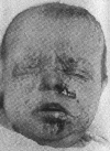
be the seat of changes. About the face, the region of the mouth,
nose, and eyelids are the sites of predilection, with frequently
resulting rhagades in these regions. A massive involvement of the
face results in a mask-like appearance. Following fissuring, there
frequently results an exudate with later crust formation. When the
scalp is involved alopecia usually results and the same may be true
when the eyelids are deeply infiltrated. The soles of the feet and
palms of the hands usually present a diffuse infiltration and appear
firm and shiny, sometimes more reddish or bluish red, at other times
a copper-red or brown.
A true paronychia, which is often accompanied by complete
destruction of the nails, is an almost constant complication in this
type of skin lesions.
The surface is either smooth or shows fissures in the uppermost
horny layers of the epidermis, which occasionally sloughs in large,
lamellous scales.
In the second type of rash lesions which are more circumscript are
noted. These lesions assume more nearly the characteristics of the
skin manifestations in acquired syphilis. The most frequent type of
lesions are macules usually circular and slightly elevated, averaging
2 to 5 mm in size. The face and the extensor surfaces of the lower
and upper extremities and more especially the hands and feet are
usually involved and they may cover the entire body, but more often
the chest and abdomen escape. At first red, they soon become darker
and assume a coppery hue. More elevated papules similar in character
and without an inflammatory base may be interspersed among the
macules. A squamous eruption is frequently seen upon the palms and
soles and small masses of scales may appear upon the surface of the
macules. The eruption may develop abruptly but more frequently it
increases progressively during a period of from one to three weeks
and under vigorous treatment disappears rapidly except for the
remaining pigmentation.
In the most severe types the bullous or pemphigoid lesions may be
superimposed upon the macular squamous syphilides or they may be
primary. They may lead to deeper ulcerations of the skin with
secondary infection and are always a source of danger to others
because of the likelihood of the presence of spirochetes in the
lesions. The possibility of the confusion of these lesions with
non-specific pemphigoid lesions which are of not infrequent
occurrence in obstetrical wards should be remembered as the latter
are especially prone to affect the premature. This latter type of
pemphigus neonatorum is probably a staphyloccus infection. Linear
fissures and mucous patches are among the most characteristic
features. On account of the fragility of the skin these rhagades
easily occur, especially on the lips, nose, about the anus and less
frequently about the eyelids. The healing of these lesions usually
results in radiating cicatrices which result in the very
characteristic "purse-string" deformity.
Umbilical Cord. -- The umbilical cord often heals slowly
and the stump has a tendency to become purulent and there is a
tendency toward infiltration about the umbilicus. A more or less
deep-seated ulcer may result which heals slowly unless constitutional
treatment is instituted or mercurials are applied locally. Hemorrhage
from the stump and secondary infection, with resulting syphilis
hemorrhagica neonatorum are likely to result.
Lymph Glands. -- Only exceptionally to they present a
characteristic generalized enlargement in the new-born syphilitic
premature. In untreated cases the lymph glands become palpable and
this may be due to luetic infection or, again, isolated groups of
glands may become involved through secondary infection and go on to
suppuration. The small size of the glands makes them difficult of
palpation, more especially in well-nourished infants.
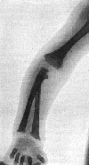
Osseous System. -- Osteochondritis syphilitica ranks next
in importance to the skin and mucous membrane lesions and splenic
tumor in the diagnosis of syphilis. Pathological changes are most
frequently seen in the long bones, the junction of the epiphysis with
the diaphysis being the seat of predilection.
These lesions are usually bilateral although occasionally only a
single lesion can be defined in the living. Involvement of the joints
is far less common. While the long cylindrical bones are the seat of
the lesions which can most easily be identified clinically, any of
the bones may be the seat of a diffuse involvement of the bony
structures or a periostitis. Such lesions are more commonly found in
cases resulting in early fetal death.
Roentgenography offers one of the best diagnostic methods
for syphilis in the fetus and new born. Shipley and his co-workers
[2] found evidence of syphilis in the osseous system
of 25 per cent of 100 white fetuses ranging from six months of
intra-uterine life to nearly term. Fifteen of these showed advanced
luetic osteochondritis. The bone lesions in syphilitic new-born
infants present characteristic lesions when there is sufficient
involvement to be evidenced in the roentgenographic plates. While any
of the bones may suffer, those most commonly involved and easiest of
study are the lower end of the femur, upper and lower ends of the
tibia, radius and ulna and the metacarpals. In their studies they
found that the fetal type of reaction and the changes before birth
were to a large extent confined to the epiphyseo-diaphyseal region,
at which points there develops an abnormal arrangement and
distribution of osseous tissue. After birth the periosteal reaction
begins, possibly because of the increased demands made on this tissue
by the increased muscular activity, and in young infants this may be
the most marked skeletal lesion. The most characteristic lesions
described by them were the following:
"The beginning of the process as shown by the
roentgen-ray picture is an intensification of the shadow cast by the
bone at the epiphyseal line. This line becomes much broader and more
homogeneous and seems to form a cap on the ends of the trabeculae of
the spongiosa (Fig. 172). This is significant of the beginning of
abnormally heavy calcification of the provisional calcified zone. It
must be remembered that while the provisional zone of calcification
in the cartilage of the normal embryonic bone is, relatively
speaking, very narrow, in may cases only one or two cells deep, in
the syphilitic bone the calcified cartilage may show on section a
width of from 0.5 to 1.5 mm.
"In other bones, in which the osteochondritis is further advanced,
it can be seen that on the marrow side of the intensified shadow of
the provisional zone there is a band-like area where the shadow is
less intense than in the rest of the bone (Fig. 170), giving an
appearance of diminished density to the region of the epiphyseal
line.
"Bones may also be seen in which the dense shadow at the
epiphyseal end of the bone is broken by the presence of one or more
small areas of rarefaction so as to give the appearance of irregular
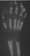
density to the end of the bone (Fig. 173).
"At other times the bone appears to end in a double line, so that
two lines of heavily calcified tissue are seen, separated from one
another by a zone in which lime salts are less heavily deposited.
This zone is a region which histological preparations show to contain
a great deal of delicate granulation tissue. This picture becomes
more and more intensified as growth goes on. The areas of dense
shadow and the fine clearer band between them grow wider and the
surfaces bounding them become more and more irregular and jagged
until the end of the bone has an irregular appearance (Fig. 171).
During the course of the disease the calcification of the infected
areas is not only abnormally heavy but also most irregular, so that
the epiphyseal border of the shadow cast by the bone has a notched,
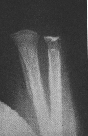
saw-toothed or serrated appearance (Fig. 175).
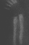
"Periostitits, when it occurs near term in the severe cases of
lues, may be present throughout the length of the bone or only at the
extremities. It is shown in roentgen-ray plates by a more or less
wide, almost homogeneous, shadow or with longitudinal striations
separated from the external surface of the cortex by a narrow clear
area which bounds the bone (Fig. 174).
"One other feature of these pictures appears worth noting. It may
be seen that in many luetic bones the cortex is separated from the
spongiosa by a very narrow clear zone which gives the cone of spongy
bone the appearance of being suspended unattached within the cortical
cavity (Fig. 175). In the roentgen-ray picture the trabeculae of the
syphilitic bone appear to be finer than those of the normal bone.
"Two other conditions which are encountered in children may give
roentgen-ray pictures which closely resemble, and in some cases are
identical with, the picture described above. Scurvy and rickets, when
the latter disease is healing under the influence of cod-liver oil
therapy, may be difficult or impossible to differentiate by
roentgenographic means from osteal syphilis of the fetal type.
Fortunately, however, in the early weeks of life neither of these
conditions need be seriously considered in diagnosticating hereditary
lues, since there is no good evidence to show that fetal rickets ever
occurs and it is agreed that scorbutus is rare before the sixth month
of life has been reached."
Liver. -- It may be stated that not less than 50 per cent
of prematurely born, syphilitic infants show a distinct enlargement
of the liver. A fair percentage of the cases show a marked increase
in size and consistency to such an extent that the abdominal
distension in its upper half is visible to the naked eye. These
latter cases are usually associated with marked jaundice, dilated
veins and an impairment of hepatic function and a high mortality.
Because of the relatively large liver of the premature new born
normally present, difficulty may be experienced in the diagnosis of a
moderate increase in size due to syphilis. It is also to be
remembered that many other factors predisposing to premature birth
have a direct influence on the size of the liver. The characteristic
pathological findings are interstitial infiltration of the connective
tissues between the acini and about the vessels. Small gummata, often
the seat of central necrosis are more exceptionally found. The
frequent involvement of the liver is readily explained by the
peculiarity of the fetal circulation. The placental blood passing
through the portal circulation by way of the umbilical veins, conveys
the spirochetes into the liver substance.
Spleen. -- Enlargement of the spleen, while usually
moderate, is one of the most important confirmatory signs but is in
itself of lesser diagnostic importance than the skin and mucous
membrane lesions. An easily palpable and hard splenic tumor in the
first three months of life, that is, before the advent of rickets in
the premature, should always be looked upon with suspicion. The
enlargement is usually due to hyperplasia of the pulp, with
occasional presence of foci of myeloid cells. Cellular infiltration
of the interstitial tissue may be present.
Respiratory System. -- The lesions of the nasal mucous
membranes have been described. Frequently there is a chronic
catarrhal laryngitis and perichondritis, with involvement of the
epiglottis. In the fetus and in infants dying soon after birth the
so-called "pneumonia alba" or "white pneumonia" is often present. In
these cases a considerable portion of the pulmonary tissue appears
whitish-gray, airless and smooth on section, due to cellular
infiltration of the interstitial tissue, filling of the alveoli and
bronchi with degenerated epithelium and proliferation of the intima
of the vessel walls. Not infrequently the pleura is the seat of small
gumma-like nodular infiltrations. Massive involvement of the lungs is
rarely compatible with life. Because of their lowered vitality,
syphilitic infants are subject to secondary bronchial and pulmonary
infection, pneumonia being a frequent cause of death.
Circulatory System. -- Most characteristic lesions are
found in the small blood vessels and careful examination shows the
presence of spirochetes in the vessel walls. These findings are most
easily demonstrated in the parenchymatous organs. The characteristic
lesions following such involvement are those of coagulation necrosis,
with secondary hemorrhages, following rupture of the vessel walls.
These lesions may result in more or less generalized or local
hemorrhagic skin lesions and those from the various mucous membranes.
Intracranial lesions frequently result from degeneration of the blood
vessels, even in the absence of trauma.
Digestive System. -- Chronic catarrhal pharyngitis is a
common early symptom which may later be followed by ulcerations of
the pharynx, tonsils and fauces. Only rarely is the stomach involved
and the lesions of the intestines which are also infrequent are
usually seen as hyperplasia of the solitary follicles and Peyer's
patches which may become necrotic and result in hemorrhages.
Scattered areas of necrosis not associated with the lymphoid tissue,
but due directly to blood vessel degeneration may be found throughout
the intestines. Peritonitis is a more frequent finding in the still
born than in viable infants. It may be of the acute type but in most
instances it is of the chronic type and may result in formation of
adhesions. Localized or generalized ascites may result.
The pancreas, thymus gland, suprarenal bodies and
thyroid gland occasionally exhibit interstitial inflammation,
gummata or other syphilitic manifestations. Small cystic formations
are frequently found in the thymus gland, usually varying in size
from 1 to 5 mm. It is a question whether they are due to arrest of
development or necrosis. Purulent material with which they are filled
contains spirochetes.
Kidneys. -- While all types of nephritis have been
described, those of greatest importance are the interstitial and
hemorrhagic. Interstitial nephritis is a serious complication because
of the danger of late secondary contraction. It is frequently
overlooked because of the absence of marked urinary findings. The
hemorrhagic types are usually associated with hemorrhages from some
of the other mucous membranes. The dangers of overmedication with
arsenic and mercury preparations, in the presence of kidney lesions
must not be overlooked. Hecker [3] states that he
has been able to demonstrate microscopic changes in 90 per cent of
his autopsies.
Nervous System. -- Involvement of the brain and its
meninges is more frequent than that of the cord. The most frequent
lesions in the still born and those dying shortly after birth is a
meningoencephalitis, involving the pia and the cortex. The pia is
infiltrated and covered by an exudate composed of plasma cells and
lymphocytes. Similar areas are seen in the cortex and the medulla may
be involved. The most frequent lesion in viable infants is a
meningitis serosa interna and externa, which is not usually noted
until after the first few weeks of life. It may develop acutely or
insidiously and usually results in a more or less marked
hydrocephalus. Because of the late development in some infants and
the early appearance of rickets in the premature it should not be
confused with megacephalus so commonly seen in the latter.
Pachymeningitis hemorrhagica less frequently seen than the former,
usually develops after the first weeks of life. Intracranial
hemorrhages are probably a more frequent cause of extra-uterine death
than is commonly supposed. Gummatous meningitis and ependymitis are
among the rare lesions. Increased intracranial pressure, as evidenced
by increased tension over the fontanelles, and which is usually
accompanied by hyperexcitability on the part of the infant, should
lead to a lumbar puncture for diagnostic purposes. Increased pressure
and an increase in the number of lymphocytes in the spinal fluid are
always suggestive but not positive evidence. A Wasserman and Lange
reaction when positive may usually be considered as conclusive
evidence. When these reactions are negative, in the presence of other
positive signs, a careful search should be made for spirochetes.
Any of the lesions of the central nervous system may result in
retarded mental and physical development.
Eyes. -- The most frequent lesions are choroiditis, optic
neuritis, iritis and parenchymatous keratitis. They are of frequent
occurrence in the still born and may develop in the first weeks of
life.
Ears. -- The organs of hearing are occasionally involved by
lesions which may be described as specific. The most common is an
involvement of the eighth nerve. Involvement of the organs of the
internal ear early in life is difficult of proof. Otitis media as
usually seen is due to a secondary infection.
Laboratory Diagnosis. -- Whenever there is a suspicion of
the presence of syphilis during pregnancy the blood of both parents
should be examined so as to give both the mother and fetus the
benefit of treatment. This will be given further consideration.
In cases in which the diagnosis has not been made before labor and
the possibility of syphilis exists the placenta should be examined
histologically and the placental cord blood should be
examined for a Wasserman reaction. It is estimated that about 50 per
cent of placentae will show more or less diffuse lesions upon
microscopic examination. Jeans and Cooke [4] found
that 57 per cent of their syphilitic infants gave a positive
Wassermann reaction on their cord blood. They found that in every
instance in which the placenta was noted as showing syphilitic
changes the infant was later found to have syphilis. While a
positive Wasserman reaction may be regarded as nearly
specific, a negative reaction must not be regarded as indicating an
absence of the disease when made during the first days or weeks of
life, as a large group of infants show little or no tendency to give
a positive Wasserman before the end of the second month of life. In
fact, some of them do not react before the end of the third or fourth
month. Negative findings in the presence of lesions or suspicion of
infection on the part of the mother should, therefore, lead to an
examination of maternal and paternal blood. The variability in the
reaction of a new born to the Wasserman test is best evidenced by the
report of one positive and one negative reaction in each of a pair of
twins by DeBuys [5] and Gerstenberger
[6].
The blood taken from the infant during the first week or two of
life shows a somewhat higher average of positive serum reaction than
examination of the placental blood taken from the same cases. In the
premature the blood can be taken from the longitudinal sinus, a scalp
vein or by a small incision in the heel. The application of the
luetin test offers serious objection in premature infants,
because of the danger of secondary infection. In full-term infants it
averages a higher percentage of positives than the Wassermann.
Demonstration of spirochetae in the open skin lesions and
bullae as well as from the scraping of the mucous membrane
ulcerations, makes the diagnosis absolute when the Treponema pallidum
is found.
While spinal and ventricular punctures are to be avoided when
possible in premature infants, examination of the cerebrospinal fluid
may be necessary when other findings are negative in the presence of
possible clinical nervous system syphilis. About 25 per cent of
new-born infants will show spinal fluid changes of sufficient
importance to have a diagnostic value. These changes consist of a
positive Wassermann, which when present, is usually associated with a
definite albumin and globulin increase. More often the cell count
reveals a moderate pleocytosis.
Prophylaxis. -- It is almost needless to say that luetic
individuals should not be permitted to marry. With the improved
methods of laboratory diagnosis of today -- luetin, Lange and
Wasserman tests -- it is now possible in a relatively high percentage
of cases to discover if an individual has a latent or active
syphilis. When there is the slightest suspicion of a specific
infection during pregnancy, the mother should be treated intensively.
This offers the only hope of preventing a similar infection of the
infant with its consequences, or of ameliorating the condition. It is
noteworthy that women with luetic histories do much better if under
treatment during pregnancy, so that prematurity and still birth may
often be avoided. In the absence of specific therapy the child,
instead of being born healthy, may show active syphilitic
manifestations or develop them later.
As in the prophylaxis of any infection of infancy, extreme care
must be exercised with reference to the sterilization of feeding and
bathing utensils and clothing.
Nursing. -- Whenever a mother bears an infant evidencing
lues, if she is at all able, she should nurse her infant. It seems
well established today that the mother is syphilitic, whether or not
her history is positive, and even in the absence of clinical
manifestations. The older controversies as to the possibility of
infection of the mother by the child and vice versa
consequently do not enter into consideration. Where an adequate
supply of milk is present it is of the utmost importance that the
premature infant be suckled. If the mother objects to nursing her
infant at the breast because of nasal and mouth lesions a shield may
be used or the milk expressed and hand fed.
Where the mother is unable to nurse her child a wet-nurse should
not be employed to suckle the child at her own breasts because of the
obvious danger of infection of the nurse. Expressed milk is, of
course very desirable.
Active Treatment. -- In syphilis neonatorum, which so
frequently is associated with serious visceral changes and so
commonly affects children born prematurely, the prognosis is in
general serious. Very commonly the infants with serious forms of
pemphigus, even with early instituted treatment and with human milk
feedings, die in the first days or weeks of life. An essentially
better prognosis is offered by the cases with maculo-papular or
papulo-bullous syphilides provided always that the internal organs
are not seriously damaged.
As soon as the diagnosis is made certain, antiluetic treatment
should be immediately instituted. Healthy infants and those free from
symptoms but born from luetic parents should be treated
prophylactically.
Certain facts already enumerated in the general care of premature
infants should be especially emphasized in the care of this same
class of infants born of syphilitic parents, even though they show no
manifestations at the time of their birth. Practically all of them
show more or less evidence of malnutrition and therefore, in this
class of infants, as in no other, is breast-feeding indicated. Every
effort should be made to stimulate the breast-milk supply on the part
of the mother because of the difficulty encountered in obtaining a
sufficient supply from other sources, as well as the danger to a
healthy wet-nurse.
In the presence of a syphilitic history or positive laboratory
findings in the parents, or the findings of clinical manifestations
in the infant a vigorous course of treatment should be instituted
without regard to the presence or absence of a Wassermann reaction.
In every case in which treatment is instituted the fetal age and
general condition of the infant must be taken into consideration and
the effect of medication, whether mercurial or arsenic preparations,
carefully noted. Early dosage with each form of medication should,
therefore, be small, however, maximum administration for the given
infant should be attained as early as possible.
Mercury Therapy. -- Three routes of administration deserve
consideration: Oral, external and intravenous.
In the treatment of older infants and children many clinicians of
large experience advocate the use of arsenic preparations as of first
importance and while mercurial preparations are considered as
absolutely necessary to effect a cure, they are given a secondary
place. In view of this tendency it is well that we recall our earlier
good results in the treatment of congenital syphilis before the
discovery of these newer preparations. It is our belief that mercury
should rank first in the treatment of syphilis in the premature and
that arsenic therapy should rank second in importance: (1) Because of
the lesser danger, and (2) because of the rapid improvement which may
be expected in a large majority of the cases. However, the dangers of
overmedication, both by mouth and injection with mercury must also
not be overlooked. These are usually evidenced by a lack of progress
on the part of the infant, diarrhea and evidences of hepatitis and
nephritis.
For internal use the favorite preparations are hydrargyrum
cum creta, 0.005 to 0.03 gm. (1/10 to 1/2 gr.), or hydrargyrum
iodidum flavum in doses of 0.002 to 0.005 gm. (1/20 to 1/10 gr.)
three times daily. It is well to begin with small doses, preferably
of the former and increase rapidly to the maximum dose in the absence
of diarrhea. in the presence of diarrhea the dose should be reduced.
The intramuscular treatment must be administered with even
greater forethought. For this purpose 0.0005 gm. (1/100 gr.) of
bichloride in 0.2 cm. (3 mm.) of distilled water or oil, for each
kilogram of body weight (2 1/2 pounds) are recommended. The
injections are to be given once or twice weekly and should be made
deep into the muscle. The gluteal muscles offer one of the best sites
for injections. The skin surface should be sterilized with a not too
concentrated tincture of iodine and the injection is made deep into
the muscle by the use of a short needle, preferably of about a 20
gauge (for oil) and 1/2 to 3/4 inch in length. Care should be used so
that none will be deposited in the subcutaneous tissues. A course of
four to eight injections, covering a period of four weeks, is
recommended, these are to be followed by a rest period of four weeks,
during which arsenic injections are given. The oral administration of
mercury should be continued throughout this period. Sublimate baths
may be successfully used in all moist forms, especially in all
exanthemata associated with vesicle formation -- 0.2 gm. (3 gr.) for
a bath of about 4 liters (1 gallon) of water.
Inunction are applicable in infants in whom the skin is not
too sensitive and are one of the best forms of treatment. In the
presence of local skin irritation it becomes necessary to stop this
form of treatment. The danger of overmedication with mercury must,
however, be borne in mind. Mercurial ointment is especially valuable
for local application to ulcerated lesions and may be applied to the
deeper seated lesions of the hands and feet by the use of mittens and
stockings. For these purposes the official mercurial ointment should
be mixed with 2 parts of lanolin. In the more mature infants it
should be carefully rubbed into the abdominal wall, axillae or thighs
and the same site used only at infrequent intervals in order that
cutaneous irritation be avoided. For a local lesion 2 per cent of
yellow oxide of mercury ointment will do. In the presence of snuffles
a 1 per cent yellow oxide of mercury ointment should be used. The
ointment is introduced into each nostril directly from a small
compressible tube. It may be necessary to carefully remove any
excessive secretions with a pledget of cotton or by washing with a
normal salt solution before applying the ointment.
It is advisable to continue mercurial treatment for at least a
year, decreasing the dose in the second six months, and repeating
three months of such therapy during the second and third years, even
in the absence of symptoms. As in treating older infants, there
should be short periods when treatment is discontinued.
Arsenic Therapy. -- It is indicated in most cases as an
adjunct to mercurial treatment. Neoarsphenamine is the preparation of
choice for use with the premature because of the fact that it can be
administered in more concentrated solution, its greater solubility
and the lack of necessity for neutralization. It can be administered
intravenously in water or by intramuscular injections in a bland oil.
The average dose is 0.01 gm. for each kilogram of body weight. The
dose should be diluted with 2 cc of sterile, freshly distilled water
for intravenous use. It is advisable to give one-half of this
quantity per kilogram for the first treatment. A course of four
intramuscular, or, when possible, preferably intravenous injections
are given at weekly intervals to be followed by a rest period of four
weeks when the treatment is to be repeated. During the period of
administration of neoarsphenamine the mercurial injections should be
discontinued but the oral administration continued.
Complications following the intramuscular injection of
neoarsphenamine, such as abscesses and infiltrations can, to a large
degree, be avoided by the use of special needles, which permit the
solution to be injected deep into the muscle. After injection, the
needle is rapidly withdrawn and a cotton pledget is pressed firmly
over the site of injection for a few minutes.
For intravenous administration the best sites are scalp veins or
the external jugular vein. For administration into the latter site
the infant should lie with the shoulders elevated and the head
extended and rotated. Only in very exceptional cases should cutting
down on a vein be practised. The longitudinal sinus route for arsenic
injection is not to be considered because of the danger of passing
through the sinus and extravasating the preparation over the brain
tissue.
The general plan of treatment should, therefore, be as follows:
One of the mercury preparations should be administered in suitable
doses three times daily per mouth, and once or twice weekly during
the first four weeks an intramuscular injection of one of the
mercurial preparations should be given. During the second month the
oral administration should be continued but the mercurial injection
should be replaced by neoarsphenamine, preferably intravenously, once
each week. Mercurial ointment as inunctions or local applications are
to be used when indicated.
This plan of treatment should be continued throughout the first
year, in the absence of toxic symptoms and at least three months of
treatment should be given during the second and third years.
Treatment should be continued for at least six months after all
evidence of activity has disappeared. This includes a negative
Wassermann test. At no time should a negative Wassermann in early
infancy be considered as sufficient evidence to interfere with the
general course of treatment as outlined.
Footnotes
[1] Am. Jour. Syph., St. Louis, 1919, No. 1, vol.
3.
[2] Shipley, P.G., Peason, J. W., Weech, A. A.,
and Greene, C. H.: Bull. Johns Hopkins Hospital, March, 1921, p. 75.
[3] Beitrag zur Histologie and Pathologie der
kongenitalen Syphilis sowie zur normalen Anatomie des Fötus und
neugeborenen, Deutsch. Arch. f. klin. Med., 1898, 61, 1.
[4] Trans. Am. Pediat. Soc., 1920, vol. 32.
[5] Jour. Obst. and Dis. Women and CHild.,
January, 1913, p. 65.
[6] Personal communication.
|

|
Fig. 167. Congenital syphilis. Secondary lesions on feet.
Lesions first appeared during fourth week.
|
|
|
Fig. 168. Congenital syphilis. Secondary lesions on face.
Lesions first appeared during fourth week.
|
|

|
Fig. 169. Congenital syphilis. Baby A. Fissures about
mouth. Large liver and spleen. Six weeks later.
|
|
|
Fig. 170. Osteochondritis syphilitica.
|
|
|
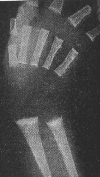
Fig. 171. Hand and forearm of human fetus to show extreme
progressive calcification of the provisional area with
irregular prolongation of the provisional calcified zone
into the area of proliferative cartilage. Note the presence
of the same lesions in the metacarpals and phalanges.
(Shipley)
|
|
|
Fig. 172. Radius and ulna from human fetus showing
beginning resorption of the area of intense calcification at
the epiphyseo-diaphyseal junction. Resorption shown by areas
of decreased intensity of shadow, each resorptive area
surrounding a small nucleus of persistent trabecular tissue.
(Shipley)
|
|
|
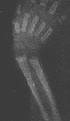
Fig. 173. Roentgen-ray picture of syphilitic
osteochondritis of the bones of the hand and forearm of a
human fetus showing a zone of rarefaction between two zones
of abnormal calcification. Note the lesion in the phalanges
and metacarpals. (Shipley)
|
|
|
Fig. 174. Syphilitic periostitis of both bones of the
forearm. Note the longitudinal striation of the thick
periosteal shadow which is nearly in contact with the shafts
of the bone. (Shipley)
|
|
|
Fig. 175. Distal end of radius and ulna. This plate shows
intense calcification of the provisional zone with
resorption areas on the marrow side of the epiphyseal line.
Both bones show syphilitic periostitis and there is
separation of the cortex from the spongiosa in the ulna.
(Shipley)
|
Return to the Hess Contents Page
Created 6/3/97 / Last modified 6/6/97
Copyright © 1998 Neonatology on the Web / webmaster@neonatology.net


