NEONATOLOGY ON THE WEB
Premature and Congenitally Diseased Infants
by Julius H. Hess, M.D.
Chapter II
Classification
For practical clinical purposes the group of infants comprising
the premature and congenitally debilitated may be classified as
follows:
- Premature infants, with no pathological changes.
- Premature infants, with pathological changes, due to:
- Constitutional disease and chronic infections in
the parents.
- Maternal factors influencing the fetal nutrition, such as
overwork, undernourishment, and acute illnesses during
pregnancy.
- Local conditions in the mother.
- Multiple pregnancies.
- Constitutional defects and congenital malformations in the
fetus.
- Infants born to parents late in life.
- Full-term infants with pathological changes due to the same
causes as those enumerated under 2.
Etiology
The occurrence of premature birth depends upon many causes, which
may be divided into those resulting in the expulsion of a healthy
premature, and those which have a damaging effect upon the product of
conception. In the first class may be included various injuries,
falls, heavy lifting, overwork or other physical exhaustion, sudden
emotional disturbances and premature rupture of the membranes, either
accidental or intentional, occurring in those conditions whose
existence does not affect the nutrition of the ovum, as in pelvic and
spinal deformity in the mother, placenta previa, etc.
Conditions in the mother requiring operative procedure not
involving the uterine cavity frequently result in premature labor
either through shock or trauma, resulting from operations, as for
ovarian conditions and uterine fibroids, or infection may be an added
danger in cholecystitis, cholelithiasis, appendicitis, ileus, and
renal operations.
The cases which fall within the second category all react to a
greater or lesser degree upon the fetus, some producing only
momentary weakness, as the milder acute infections, others causing a
weakened physical condition as a result of their long-continued
action upon the nutrition and development of the fetus.
The most frequent causes are the chronic infections.
Syphilis plays the leading role, and is estimated as being a
factor in from 50 to 80 per cent of all cases of repeated premature
expulsion of the fetus, while Lesage and Kouriansky
[1] state that syphilis is a factor in the causation
of congenital debility of the full-term in 25 to 35 per cent. If the
luetic infection is recent, abortion is the rule; but as the
infection becomes older, the succeeding pregnancies terminate later
and later until a living child with or without manifestations of the
disease is born, usually prematurely.
Chronic nephritis is one of the most frequent causes of
spontaneous premature labor, and the offspring of these mothers are
often puny, due, either to the systemic affect on the mother, or
resulting from impaired nutrition of the fetus due to placental
hemorrhages and infarcts. Nephritis in the mother is also one of the
most frequent indications for the induction of premature labor.
Pulmonary tuberculosis is less frequently the cause of
premature labor, but the children, even at full-term, are often small
and weak. Tuberculosis of other organs and tissues influences the
fetus in proportion to the nutritional effect upon the mother or,
again when involving the vertebral column or hip-joints may by their
resulting deformities require premature induction of labor.
Congenital tuberculosis is very rare, but does occur. In the majority
of cases, not the disease per se, but the predisposition is
inherited.
Premature birth occurs in 30 or 35 per cent of the cases of broken
compensation in heart disease. The premature infants are, in
these cases, often imperfectly nourished as a result of the poor
aeration of the mother's blood. Exophthalmic goiter is
occasionally the cause of premature emptying of the uterus. If
chronic dyspnea exists, as a result of laryngeal or tracheal stenosis
from pressure, the development of the fetus will necessarily be
retarded.
Any of the acute infectious diseases may be responsible for
the termination of pregnancy before the end of term. Pneumonia,
influenza, typhoid fever, malaria, diphtheria, scarlet fever,
measles, small-pox, Asiatic cholera and bubonic plague -- all have a
deleterious effect on the continuance of pregnancy. Premature labor
is very common in pneumonia and influenza, being more frequent in
late pregnancy.
Of local conditions, diseases of the decidua or
endometrium, gonorrheal infection and malpositions of the uterus
frequently result in premature labor, but usually before the fetus is
viable. Anomalous positions of the fetus in utero may be
responsible for the premature expulsion of the uterine contents.
The occurrence of multiple pregnancy is a fruitful source
of premature labor. About 70 per cent of twin pregnancies terminate
prematurely and the length of practically all triplet and quadruplet
gestations is considerably shortened in most cases due to lack of
room in the uterine cavity. Miller's [2] figures are
slightly smaller. He states that of 3380 plural births, 2040, or 60
per cent, were premature, and had a body weight of less than 2500
gm., and a length under 45 cm. Even when mature, twins are usually
small and of low body weight. This, of course, is even more true of
triple pregnancies, the reserve strength possessed by the mother not
being sufficient to allow three fetuses to reach their normal
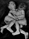
development. Again in the presence of several fetuses the growth may
proceed unequivocally so that one may be born with unimpaired
vitality, and the others with greatly diminished strength
(Fig. 9).
Faulty nutrition of the fetus, such as is found in maternal
overwork or from lack of sufficient food, as well as that due to
wasting diseases, the blood dyscrasias (pernicious anemia and
leukemia), and intoxication from alcohol (acute and chronic),
phosphorus, arsenic, mercury, or lead may -- any one of them -- cause
either an early termination of pregnancy or so serious a lowering of
nutrition of the fetus that the vitality at birth may be greatly
impaired. In addition, congenital malformations in the fetus
sometimes bring on premature birth. In diabetes prematurity is
not infrequent, and the infants may show glycosuria.
Infants born to parents late in life are often born prematurely,
perhaps because of the factor of undernourishment. This is also the
case in prematures born of women who have had numerous pregnancies,
at short intervals.
Finally, habitual miscarriage, without evident cause, resulting in
the interruption of successive pregnancies, not infrequently at about
the same stage, is not rare. The author has records of several such
women without a history of syphilis or other constitutional disease,
and in whom uterine deformity is not demonstrable.
The frequency of premature labors varies greatly in
different clinics. Rommel [3] quotes the following
figures from various clinics, noting the number of infants under 2500
gm. in weight and below 45 cm. in length.
|
Miller
|
5.0 per cent
|
Orphan Asylum
|
Moscow
|
|
Von Winckel
|
13.3 per cent
|
Maternity
|
Munich
|
|
Fehling
|
25.0 per cent
|
Maternity
|
Halle
|
|
Budin
|
10.7 per cent
|
Clinique Tarnier
|
Paris
|
|
Pinard
|
15.4 per cent
|
Clinique Baudelocque
|
Paris
|
It is stated that the percentage of premature births is greater
during the colder months of the year.
Footnotes
[1] Congenital Debility and Atrophy, Nourrisson,
Paris, July, 1919, No. 4, 7, 193.
[2] Peculiarities of the Disease of the Premature
Infant, Jahrb. f. Khlk., 1886, 25, 129.
[3] Quoted from Pfaundler and Schlossman Handb. f.
Kinderh., Leipzig, 1901.
|

|
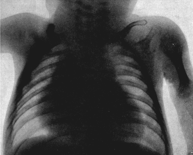
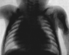
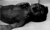
Fig. 1. Case of congenital goiter.
|
|
|
Fig. 2. Case of congenital thymus (atrophy of gland
following two exposures to roentgen ray).
|
|
|
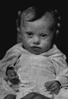
Fig. 3. Mongolian idiot.
|
|

|
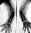
Fig. 4. Chondrodystrophia.
|
|
|
Fig. 5. Chondrodystrophia.
|
|
|
Fig. 6. Cretinism.
|
|

|
Fig. 7. Dyspituitarism.
|
|
|
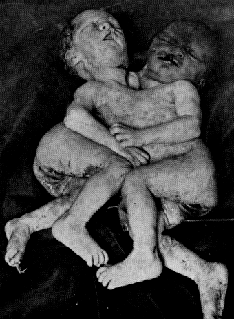
Fig. 8. Case of Siamese twins. Thoracopagus tetrabrachius
tetrapus. (From the service of Dr. Ludwig Simon, Michael
Reese Hospital, Chicago.)
|
|

|
Fig. 9. Triplets.
|
Return to the Hess Contents Page
Created 4/18/97 / Last modified 4/18/97
Copyright © 1998 Neonatology on the Web / webmaster@neonatology.net