by Julius H. Hess, M.D.
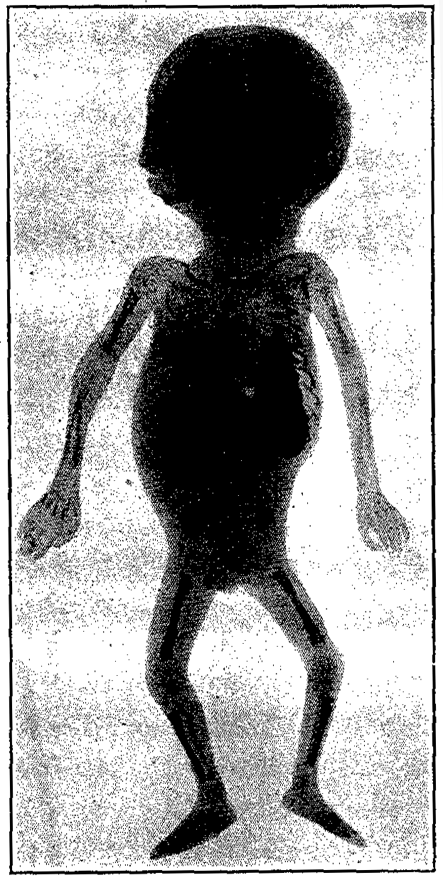
The appearance and characteristics of the healthy premature child vary with the fetal age at the time of birth. With a lengthening of the period of gestation, the` distinctive characteristics of the fetus become less and less marked until it becomes impossible to differentiate the slightly premature from the full-term infant. All the distinguishing features of the premature may also be, found in the congenitally diseased full-term infants, and as there may be all degrees of prematurity, so we also find all stages of development between the extremes of functional and anatomical inferiority on the one hand and the normal constitution on the other. Both the premature and the debilitated infant may exhibit the following features in varying degrees.
The body is usually small and puny, though in some instances the infant may be of a considerable size, yet with a very imperfect development of its internal organs.
The weight is low, varying from amounts approximating 700 gm. (1 1/2 lbs.) to 2500. gm. (5 1/2 lbs.) in the viable. The latter figure may be exceeded in infants nearing maturity, and by some of the full-term weaklings, but will serve as a fair maximum.
The skin is soft and usually of a vivid red color. The epidermis is thin and the blood vessels are easily seen.
The skin frequently hangs in folds. The adipose tissue is scant, the features are angular and the face looks old.
Lanugo is plentiful, especially upon the extensor surfaces of the extremities.
The skull is round or ovoid in contradistinction to the usually markedly dolichocephalic skull of the full-term new-born. The fontanelles are large and the sutures prominent.
The nose, exhibits many small comedones. The ears are soft and small and hug the skull.
The nails have scarcely reached the ends of the fingers even in the larger infants, while in the smaller they may be very poorly developed. The cry is feeble, monotonous and whining.
The infant lies in a deep sleep, and must be aroused for its feedings. Efforts at suction are weak or absent. All movements are slow, functions are sluggish and the child shows a remarkable degree of muscular inertia.
The temperature has a very decided tendency to remain below-normal and is inclined to be irregular in character.
The urine is usually scanty.
The bowels are sluggish and constipation is the rule. Early and intense jaundice is common.
These are the principal findings which are to be seen on superficial examination. A more critical review of these various characteristics follows. It must be remembered that any of these symptoms may vary in different individuals of the same age, depending upon the cause of prematurity, and upon the condition of health present in both the mother and the child. With increasing age, the characteristics become less marked, until the picture eventually merges into that of the full-term infant.
The determination of the exact age of the infant prematurely born is a matter of considerable difficulty. The information furnished by the mother as to the time of her last menstrual period, or as to the time when life was first felt, gives an entirely insufficient approximation of the probable date of confinement, and errors of a month or even more are not rare. In institutions for foundlings all data is, as a rule, absent, and other methods for determining the infant's fetal age must be relied upon. The weight of the infant is of uncertain value also, as an infant of 1500 gm. weight may be the product of a pregnancy of seven months in a healthy woman, while one of the same or less weight may be the eighth-month offspring of an albuminuric or syphilitic mother. The body measurements also vary materially with the individual. The degree of development of the osseous system is of great value in determining the anatomical development, and indirectly the condition of the bones acts as a guide to physiological development, even though they do not give absolute data as to age. Body measurements and osseous development are fully discussed later under their respective headings.
More important than a determination of the approximate term of pregnancy or a consideration of the size of the infant, at least in those infants born but a few weeks before the natural termination of the period, is a history of syphilis, tuberculosis, traumata, or other causes, operating in the mother and responsible for the early emptying of the uterus.
His gives the following description of the developmental features of the fetus at varying ages:
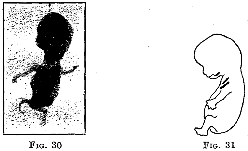
Fifth Lunar Month (112 to 140 days).-Head about the size of hen's egg; the skin is red and shows some fat deposit. The scalp shows indications of hair, the body is covered with lanugo, the nails can be distinguished, the eyelids remain closed. The fetus rarely lives over five to ten minutes, making feeble attempts at respiration. The heart-beats may be strong.
Sixth Lunar Month (140 to 168 days).-The body shows increased fat deposits, though still lean, the skin being wrinkled. The eyelids are separated and eyebrows and lashes may be seen. The infant may live for several hours. The respiratory and digestive organs are underdeveloped, respirations being superficial and digestion practically impossible.
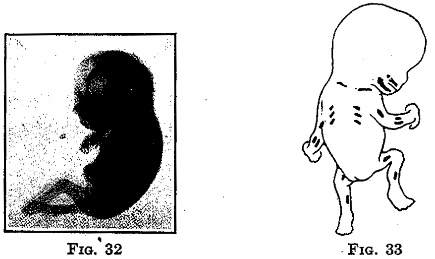
Seventh Lunar Month (168 to 196 days).-The infant has an aged appearance but the wrinkles are filling out. The eyes are open. The cry is a weak whine or grunt. Few of these infants born during the twenty-fifth and twenty-sixth weeks survive, and when they do are usually hydrocephalic, paralytic and dwarfed. Those of the twenty-seventh and twenty-eighth weeks are far more promising.
Eighth Lunar Month (196 to 224 days).-The infant is beginning to fill out, many of the wrinkles having disappeared. The bones of the head are soft and flexible. Ossification begins in the lower epiphysis of the femur. The testicles are often in the scrotum. The cry is stronger, though it may still be very weak. Under proper conditions many of these infants survive.
Ninth Lunar Month (224 to 252 days). -Panniculus adiposus develops. The wrinkles smooth out and the limbs become rounded. The lanugo begins to disappear, and the nails are at the tips of the fingers. Respiratory, circulatory and digestive organs are capable of carrying on the. body functions.
Tenth Lunar Month (252 to 280 days).-The general body functions improve during this month and at the end of this period development is complete.
Infants born at full-term weigh on the average from 3000 to 3500 gm. The dividing line between the premature and full-term infant has been generally placed at 2500 gm. If under that figure they may be considered below par as far as concerns the strength and ability to overcome the forces which assail them on every hand. The weight of the premature varies even within greater limits than that of the full-term infant, and as one may see a child below 2500 gm, so also there are prematures with a weight above this limit.
The weight depends upon the cause of the premature birth and upon the age of the child. Those born of mothers afflicted with nephritis, tuberculosis, or other wasting diseases, and infants showing active syphilis, are usually considerably smaller than the same aged infants of healthy parents. Diseases and abnormal location of the placenta also restrict the growth of the fetus. The infant in placenta previa is often undersized, even when born at term. Multiparity may predispose to undersize.
His, in a comparison of the fetal weight and length with the age, made the following table:
|
|
Weight |
Length |
|
16 to 20 weeks |
250 to 280 gms |
17 to 26 cm |
|
20 to 24 weeks |
645 to 1000 gms |
28 to 34 cm |
|
24 to 28 weeks |
1000 to 1220 gms |
35 to 38 cm |
|
28 to 32 weeks |
1220 to 1600 gms |
39 to 43 cm |
|
32 to 36 weeks |
1600 to 2500 gms |
46 to 48 cm |
|
36 to 40 weeks |
2500 to 3100 cm |
48 to 50 cm |
THE AVERAGE LENGTHS IN CENTIMETERS OF NORMAL
FETUSES AS GIVEN BY DIFFERENT OBSERVERS.
[The length for the first two months represents
the
measurement from the vertex to the buttocks;
all the other measurements are from vertex to sole.]
|
Lunar Months |
Mall |
Von Winckel |
De Lee |
Lambertz |
Ahlfeld |
Schroeder |
|
1st |
0.25 |
|
0.75-0.9 |
|
|
|
|
2nd |
0.55-3.0 |
0.9-2.5 |
2.5 |
|
|
|
|
3rd |
4.1-9.8 |
7-9 |
7-9 |
6-11 |
|
|
|
4th |
11.7-18.0 |
10-17 |
10-17 |
11-17 |
|
|
|
5th |
19.8-25.0 |
18-27 |
17-26 |
17-28 |
|
|
|
7th |
33.1-37.1 |
35-38 |
35-38 |
35-38 |
36-40 |
|
|
8th |
38.4-42.5 |
40-43 |
45 |
38-42 |
40-43 |
41.3 |
|
9th |
43.6-47.0 |
46-48 |
40-48 |
42-45 |
46-48 |
44.6 |
|
10th |
48.4-50 |
48-50 |
48-50 |
45-52 |
48-50 |
46.0 |
The weight and length as compared to the fetal age is shown in the following table from Oberwarth, which gives the average length also:
|
Fetal age |
Weight |
Length |
|
26 weeks |
330 to 1041 gms |
28.0 to 37.0 cm |
|
28 weeks |
995 to 1408 gms |
36.3 to 37.5 cm |
|
30 weeks |
797 to 1700 gms |
33.1 to 41.3 cm |
|
32 weeks |
1868 to 1964 gms |
42.0 to 42.7 cm |
|
34 weeks |
1286 to 2213 gms |
39.0 to 47.0 cm |
|
36 weeks |
2424 to 2700 gms |
46.1 to 48.0 cm |
These compare favorably with those given by Ahlfeld and Hecker.
|
Fetal age |
Weight |
Length |
|
27 weeks |
1140 gms |
36.3 cm |
|
29 weeks |
1575 gms |
39.6 cm |
|
31 weeks |
1975 gms |
42.7 cm |
|
33 weeks |
2100 gm |
43.9 cm |
|
35 weeks |
2750 gms |
47.3 cm |
|
37 weeks |
2875 gms |
48.3 cm |
Potel and Hahn's figures do not include the length.
|
Fetal age |
Weight |
|
27 weeks |
995 to 1146 gms |
|
29 weeks |
1540 to 1700 gms |
|
31 weeks |
1881 to 1964 gms |
|
33 weeks |
2150 to 2213 gms |
|
35 weeks |
2400 to 2700 gms |
The following small group taken from my cases give the age of the fetus as computed from. the date of the last menstruation. That this is an unreliable method may be recognized by noting the variation in figures in Cases 2, 3, 11, 13, 14 and 15. We therefore, place. little reliance on the mother's estimate as to the date of conception.
|
|
|
|
|
|
||||
|
Case |
Fetal age, weeks |
Weight, gm |
Length, cm |
O. F. |
Bi. P. |
Bi. T. |
Oc. M. |
S. O. B. |
|
1 |
21 |
700 |
30.0 |
7.5 |
5.5 |
4.5 |
9.0 |
7.5 |
|
2 |
22 |
1015 |
37.9 |
7.5 |
6.5 |
6.0 |
9.0 |
7.5 |
|
3 |
27 |
1690 |
40.9 |
9.0 |
8.0 |
6.5 |
11.0 |
7.5 |
|
4 |
29 |
1449 |
|
8.0 |
7.0 |
7.0 |
8.0 |
7.0 |
|
5 |
31 |
1175 |
37.5 |
9.0 |
7.0 |
6.0 |
11.0 |
8.0 |
|
6 |
32 |
1380 |
34.0 |
9.0 |
8.0 |
7.0 |
11.0 |
7.0 |
|
7 |
32 |
2040 |
45.0 |
11.5 |
8.5 |
7.5 |
13.0 |
9.5 |
|
8 |
33 |
1175 |
44.0 |
9.0 |
7.0 |
6.0 |
11.0 |
8.0 |
|
9 |
33 |
2110 |
45.0 |
10.0 |
8.0 |
6.0 |
12.0 |
8.0 |
|
10 |
38 |
3625 |
50.0 |
11.0 |
9.5 |
8.0 |
13.25 |
9.5 |
|
11 |
39 |
1610 |
41.5 |
10.0 |
7.75 |
6.25 |
11.75 |
8.5 |
|
12 |
39 |
3260 |
49.0 |
11.5 |
9.5 |
8.5 |
13.5 |
9.75 |
|
13 |
40 |
1370 |
38.0 |
9.0 |
7.0 |
6.0 |
10.0 |
8.0 |
|
14 |
41 |
1570 |
35.0 |
11.0 |
8.0 |
7.5 |
11.5 |
7.0 |
|
15 |
41 |
1810 |
38.5 |
10.0 |
8.0 |
7.5 |
12.5 |
8.5 |
In contrast with these measurements of the diameters of the head in prematures, the average measurements of the skull in a mature new born are noted as follows by Schauta.
1. Diameter suboccipito-bregmaticus (from the posterior edge of the great occipital foramen to the anterior angle of the great fontanelle), 9 cm.
2: Diameter fronto-occipitalis (from glabella to the occipital protuberance), 11 cm.
3. Diameter mento-occipitalis (from the point of the chin to the farthest point of the occiput), 13 cm.
4. Diameter verticalis (from the vertex to the base of the skull), 9.5 cm.
5. Diameter biparietalis (between the parietal tuberosities), 9 cm.
6. Diameter bitemporalis (between the farthest point of both coronary sutures), 8 cm.
Parents short in stature or small in build may have children who do not weigh over 2000 gm. or measure over 45 cm in length, and yet who are neither premature nor congenitally weak.
It does not do to estimate the vitality of these infants from a consideration of their birth weight. Many of them born at or near term have a normal weight, yet they do not survive. On the other hand, infants of considerably less weight may present evidence of great vitality, a lusty cry and take nourishment with avidity. According to our experience the condition of the turgor of the prematurely born infants is of much more importance than all these. Flabby prematures with a poor turgor and a poor tonus are usually not viable. Prematures with a good turgor and a good tonus even with a low weight commonly survive.
In addition to the variations in weight and length, the premature shows variations in other measurements.
Other Measurements of the Fetus.-Von Winckell regards the circumference of the head as of importance for the diagnosis of the age of the fetus and gives the following figures:
|
4th month |
10-14 cm |
|
5th month |
13-18 cm |
|
6th month |
19-24 cm |
|
7th month |
23-28 cm |
|
8th month |
25-30 cm |
|
9th month |
29-33 cm |
|
10th month |
32-37 cm |
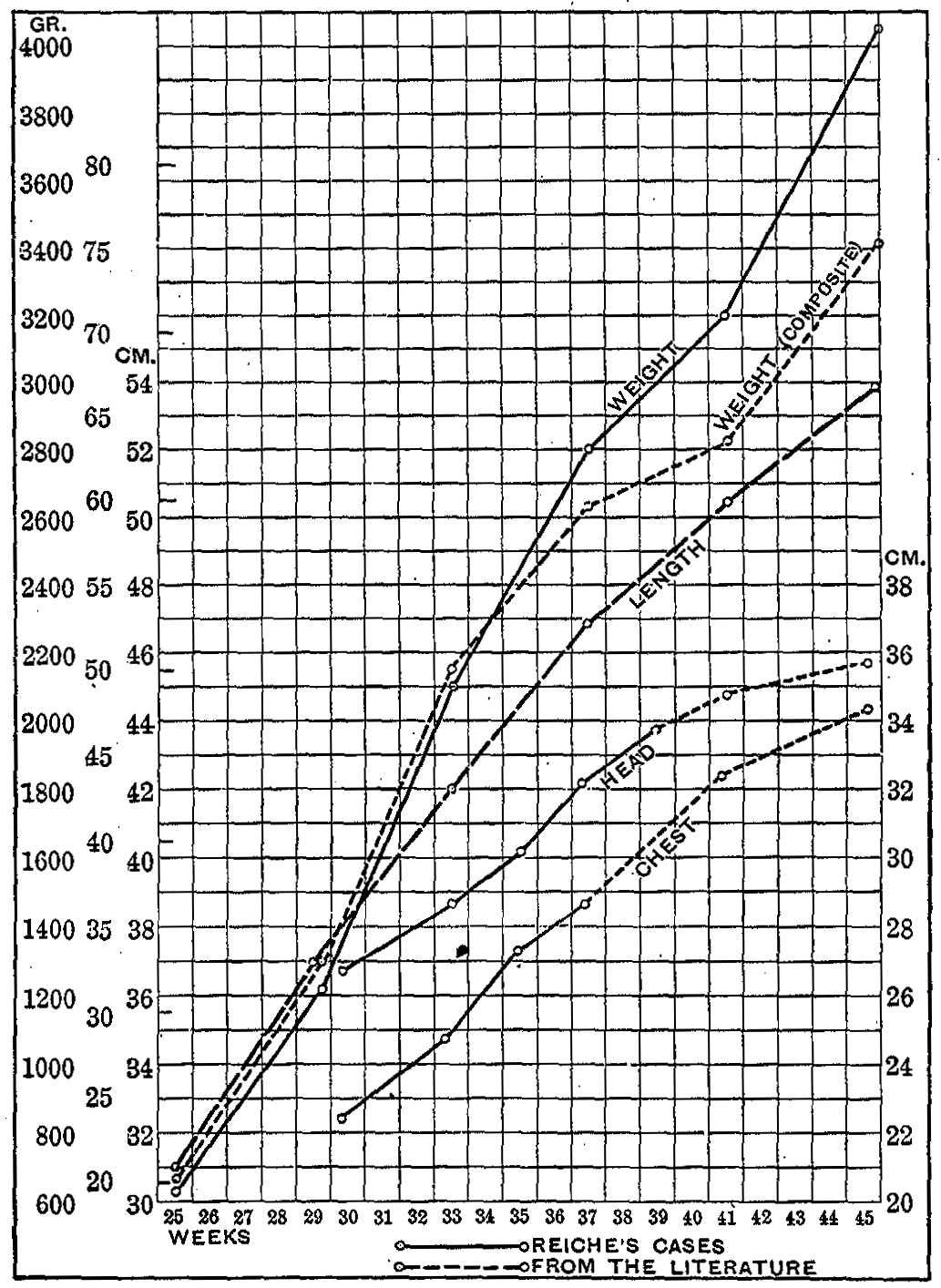
Reiche reports the following comparative body measurements:
|
|
|
||
|
|
|
|
|
|
Length of the body |
|
|
|
|
Circumference of chest |
|
|
|
|
Circumference of head |
|
|
|
|
|
|
||
|
|
|
|
|
|
Length of the body |
|
|
|
|
Circumference of chest |
|
|
|
|
Circumference of head |
|
|
|
|
|
|
||
|
|
|
|
|
|
Length of the body |
|
|
|
|
Circumference of chest |
|
|
|
|
Circumference of head |
|
|
|
|
|
|
|
|
|
|
|
||
|
|
|
|
|
|
Length of the body |
|
|
|
|
Circumference of chest |
|
|
|
|
Circumference of head |
|
|
|
These figures show a gradual and steady increase of the weight and the chest and head measurements, up to the time of maturity, when they should average 3200 gm in weight, 50.5 cm. in length, with a chest circumference of 32.9 to 33.8 cm. and a head circumference of 34.5 cm.
We see in the eighth to the tenth month an abrupt rise of the curve of chest circumference, the curve flattening somewhat soon after birth. This increase in the circumference of the chest in the last fetal months is considerably higher than that of a mature child during the first months after birth. In the latter the circumference of the chest increases from 32.5 to 37.2 at the end of the third month to 41 at the end of the sixth month, therefore in the first six months of life approximately about as much as in the last three fetal months.
In the curve of the growth of the skull the flattening appears even somewhat earlier. The ratio, however, between the growth of the skull in the last three fetal months and that in the first six months of life is the same as in the circumference of the chest. Also the circumference of the head grows absolutely and relatively considerably more in the last fetal months than in the first six months of life.
A proof for the correctness of these figures Reichel finds in the fact that the corresponding figures are considerably lower in children who die shortly after birth. They are premature weaklings whose intra-uterine development in spite of sufficient body weight did not attain such a degree that it might be completed in the extra-uterine life.
The corresponding figures are, as follows:
|
|
|
||
|
|
|
|
|
|
Length of the body |
|
|
|
|
Circumference of chest |
|
|
|
|
Circumference of head |
|
|
|
|
|
|
||
|
|
|
|
|
|
Length of the body |
|
|
|
|
Circumference of chest |
|
|
|
|
Circumference of head |
|
|
|
|
|
|
||
|
|
|
|
|
|
Length of the body |
|
|
|
|
Circumference of chest |
|
|
|
|
Circumference of head |
|
|
|
From these figures Reiche concludes that in premature weaklings the length of the body does not vary greatly from that of healthy children, but on the other hand the measurements of the circumference of the chest and of the circumference of the head are considerably smaller.
Ylppo recently studied the relation of the chest circumference to that of the head in prematures and full-term infants. He found that at birth the circumference of the bead is greater than that of the chest, and the greater the prematurity the more marked is the relative disproportion between the head and chest circumferences. These facts are borne out by his table:
|
Weight of infants, grams |
Number |
Circumference of head |
Circumference of chest |
Breast circumference, per cent of head circumference |
|
Under 1000 |
16 |
25.0 |
20.8 |
83.2 |
|
1001-1500 |
78 |
31.8 |
24.5 |
77.0 |
|
1501-2000 |
75 |
30.0 |
26.3 |
87.7 |
|
2001-2500 |
74 |
32.3 |
29.5 |
91.3 |
|
New born |
100 |
33.5 |
31.0 |
92.5 |
In comparison with the preceding tables on prematures we note the conclusions drawn by von Reuss from his own work and the tabulations compiled by Weissenberg on the mature new-born infant.
|
|
|
|
||||
|
Body measurement |
Min |
Max |
Average |
Min |
Max |
Average |
|
Body length |
47.5 |
54.0 |
50.8 |
43.5 |
53.0 |
50.0 |
|
Span of arms |
45.0 |
52.0 |
48.6 |
42.0 |
52.0 |
48.0 |
|
Vertex-shoulder |
11.5 |
13.5 |
12.4 |
10.5 |
13.5 |
12.1 |
|
Sitting-height |
31.2 |
36.5 |
33.8 |
30.0 |
36.4 |
33.3 |
|
Breadth of shoulders |
9.0 |
12.2 |
10.7 |
9.0 |
12.0 |
10.4 |
|
Breadth of hips |
7.0 |
8.7 |
7.8 |
6.8 |
8.3 |
7.7 |
|
Circumference of head |
30.5 |
35.5 |
32.7 |
29.0 |
35.0 |
32.6 |
|
Girth of chest |
25.5 |
32.0 |
28.2 |
25.0 |
32.0 |
28.5 |
|
Length of trunk |
19.5 |
24.0 |
21.4 |
19.0 |
24.0 |
21.2 |
|
Length of arm |
19.5 |
23.5 |
21.4 |
18.5 |
22.5 |
21.0 |
|
Length of leg |
18.0 |
22.2 |
20.5 |
17.0 |
21.8 |
20.3 |
|
Length of hand |
5.8 |
7.0 |
6.4 |
5.8 |
7.5 |
6.4 |
|
Length of foot |
7.3 |
8.3 |
7.8 |
6.5 |
8.3 |
7.8 |
The peculiarities of the proportions of the body characteristic of the full-term new born consist therefore of the following: Not only the sitting height, but also the height of the trunk proper is greater than the leg. The length of the trunk proper is greater than that of the arm. The arm is longer than the leg. The circumference of the head is usually greater than that of the chest. Occasionally the circumference of the head and chest are equal; in strongly built infants the circumference of the chest often exceeds that-of the head. The body length approximates 47 to 54 cm. and errors in statements of length result because of the lack of consideration for the deformity of the skull and caput succedaneum (von Reuss).
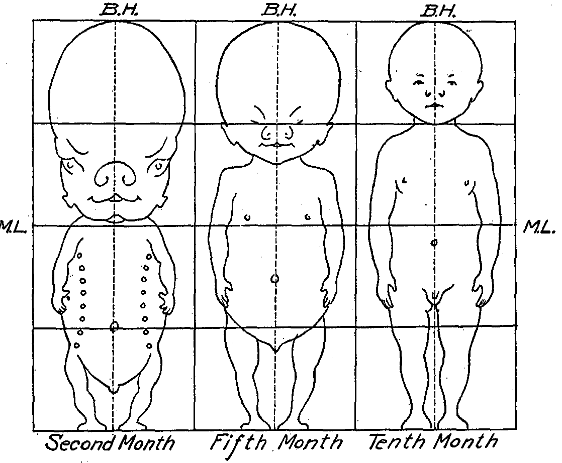
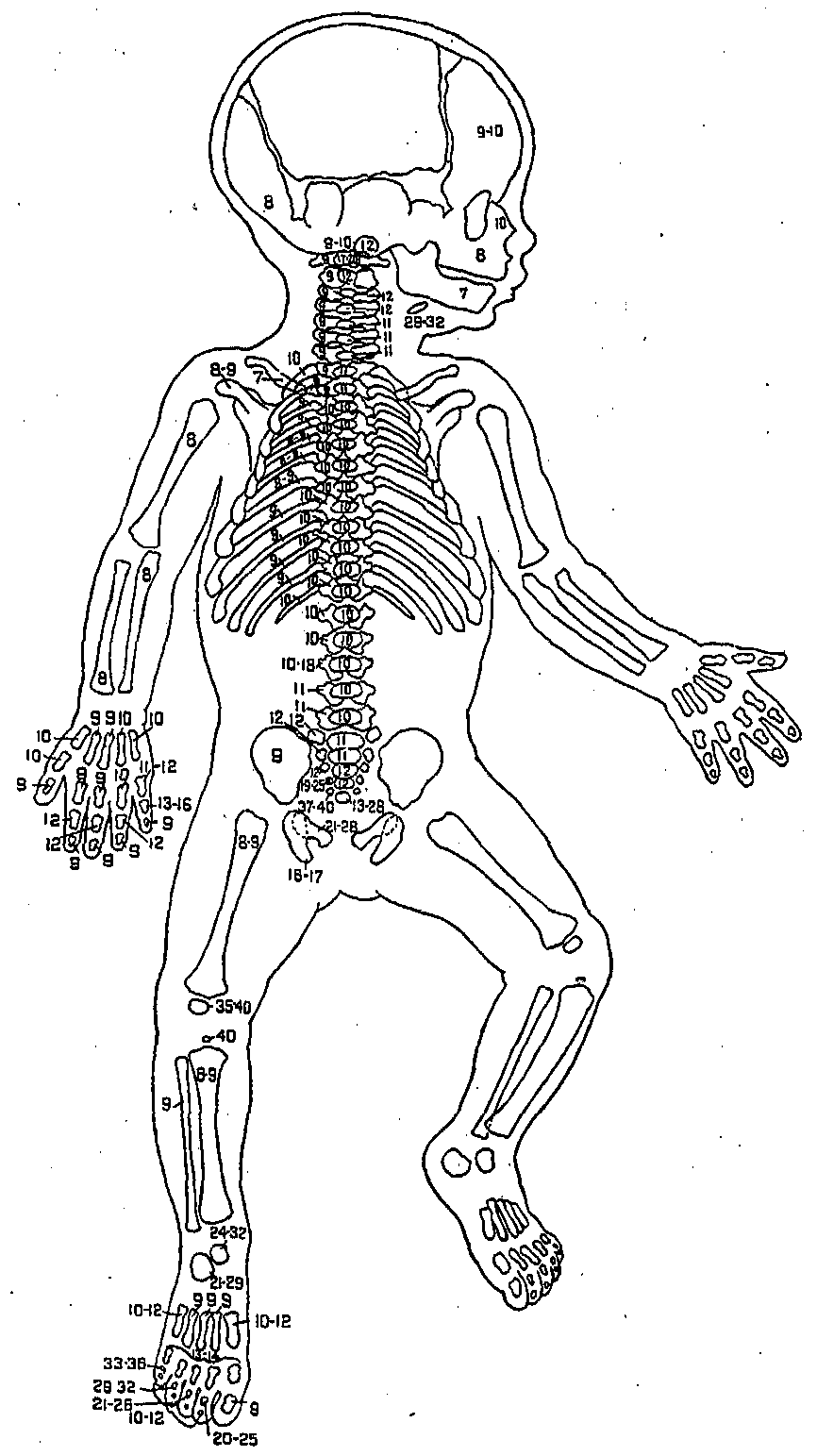
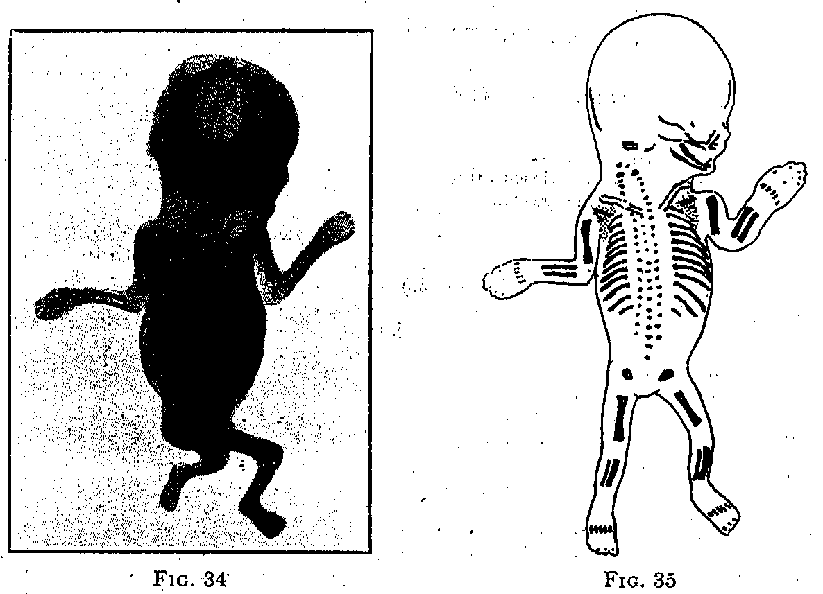
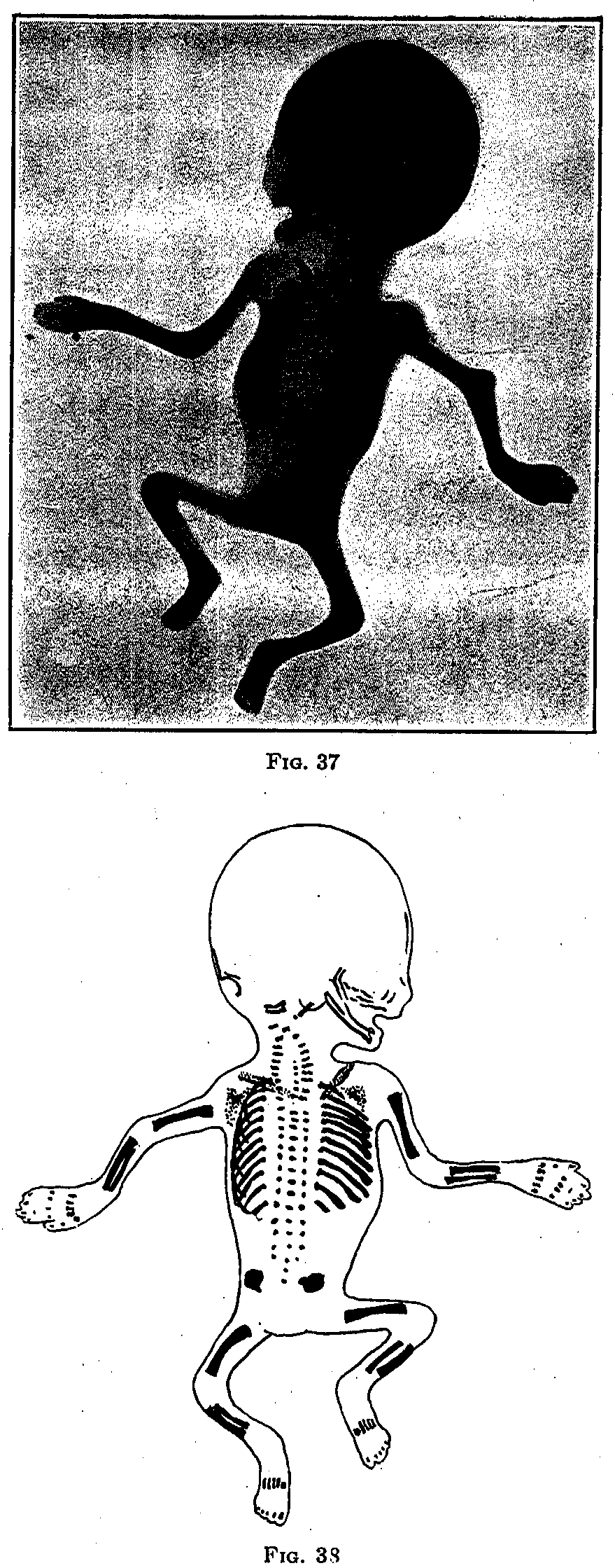
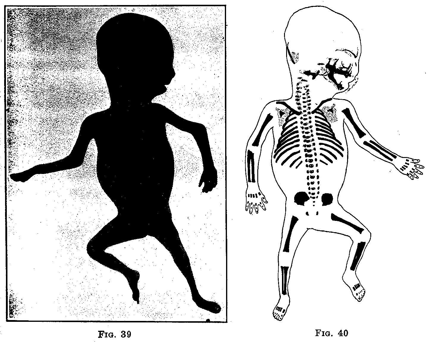
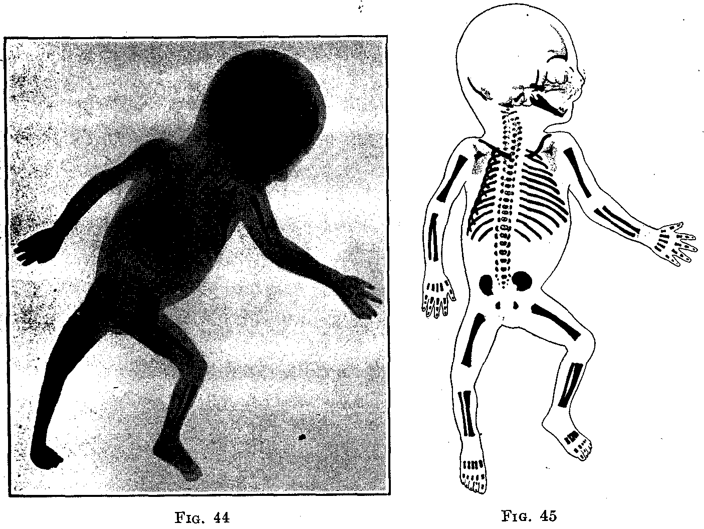
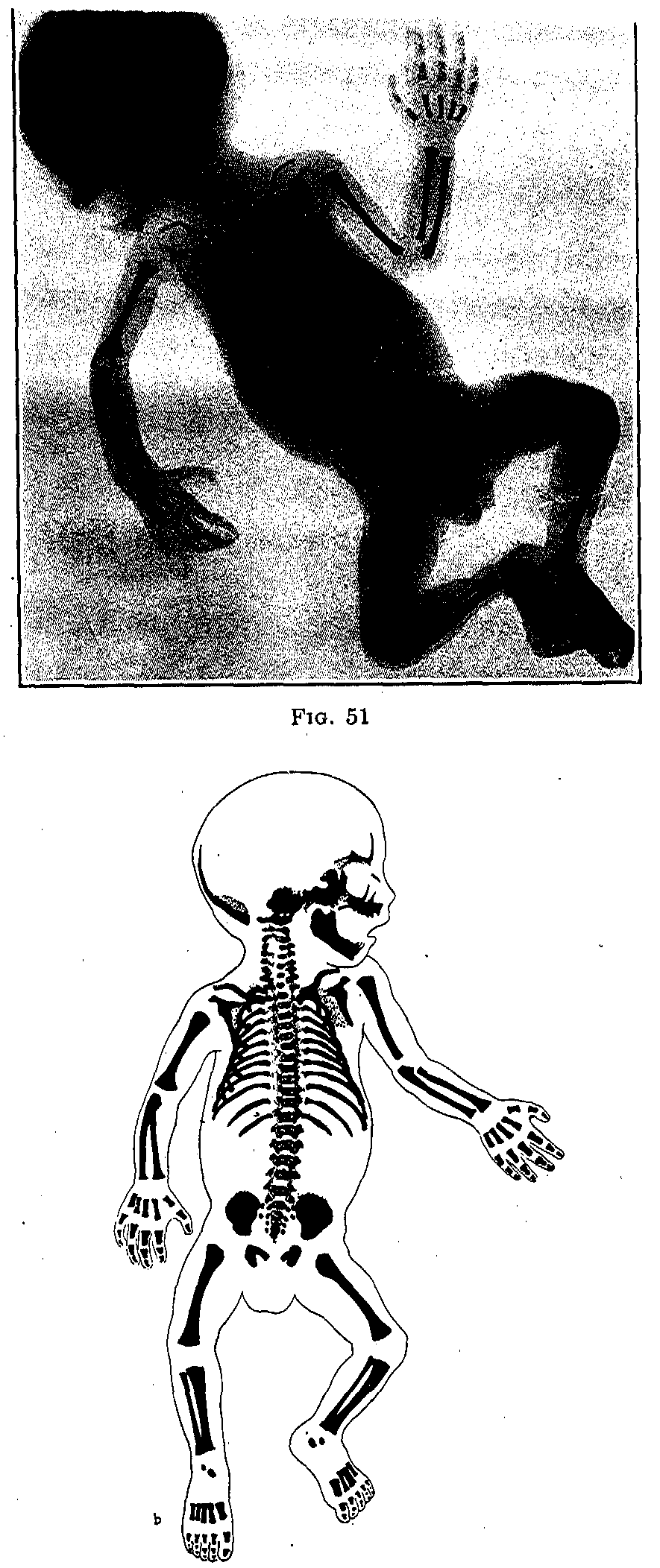
Jaschkem in a recent study of the premature and debilitated child, came to the conclusion that there was less variability in certain relations between measurements of the body than was commonly thought. "In immature infants the fronto-occipital circumference of the head always is greater than the circumference of the shoulders (Frank and others), while in mature infants the opposite is true; also the proportion between the height of the head and the height of the body (Stratz) is disturbed since the height of the head is greater than one-fourth of the length of the body; this is due especially to relatively shorter legs" (Fig. 11).
Gundobin, studying the average weight of the inner organs of the mature new born in grams, noted the following:
|
Brain |
389-354.5 |
|
Heart |
17.24-16.5 |
|
Lungs |
57 (Lt. 25;Rt. 32) |
|
Liver |
120-130 |
|
Pancreas |
2.63 |
|
Spleen |
7.2 |
|
Kidneys |
11-12 |
|
Suprarenals |
2.5 |
|
Testicles |
0.2 |
|
Epididymes |
0.12 |
|
Ovaries |
0.2 |
|
Thyroid |
1.6 (Max 2.8; Min 1.3) |
|
Thymus |
11.7 |
In contrast with these figures, we may quote from the anatomical studies of Ylppo on premature infants.
|
|
|
|
||
|
Age |
Number of cases |
Of body |
Of entire brain |
Ratio of brain to body weight |
|
Fetus of eight months |
3 |
2440 |
248 |
1 to 10 |
|
Newly born |
3 |
2785 |
389 |
1 to 7.2 |
|
1 month |
3 |
3860 |
517 |
1 to 7.5 |
|
2 months |
5 |
4400 |
538 |
1 to 8.2 |
|
3 months |
5 |
4480 |
555 |
1 to 8.1 |
|
4 months |
5 |
4890 |
568 |
1 to 8.6 |
|
5 months |
5 |
5614 |
632 |
1 to 8.9 |
|
6 months |
5 |
6035 |
668 |
1 to 9.0 |
|
7 months |
3 |
6560 |
702 |
1 to 9.3 |
|
8 months |
3 |
6460 |
768 |
1 to 8.4 |
Ylppo found several instances in which the large brain weight seemed to be out of proportion to the figures of other observers. His studies led him to believe that the brain of the premature (even the smallest) grows at the same rate as if the fetus were in utero and that it develops in extra-uterine life after certain given laws of Nature; thus, the small body weight having relatively little to do with the brain. In these cases of marked disproportion he found that when one compares the absolute age of the premature, from the time of conception, with that of a normal infant, it is seen that the brain weight of the two compare favorably. His conclusions were that the size of the brain has nothing to do with a hydrocephalic process, since it is not explained by an abnormal water content, and that the "megacephaly" of prematures is a physiological process.
Tonsils.-In prematures there appears at the site of the palatine tonsils only one or two small cavities. Only after four to five months does a glandular structure appear.
Thyroid Gland.-This is very small, but it has a very rich blood supply. In one case of a seven-months premature Ylppo observed an enlargement of the thyroid (1.5 gm.): weight of infant, 1270 gm.; length, 44 cm. Microscopically there were large quantities of colloid in the center of the follicles, but no hemorrhages or evidence of degenerative changes.
Thymus Gland.-In prematures of 1000 to 2000 gm it is between 1 and 3 gm., while in full-terms it may be as much as 20 gm. Gundobin estimated it in prematures of similar weight as on the average of 2.5 gm.
Heart.-The heart on the average is from 0.5 to 0.75 per cent of the body weight of prematures. In those from 900 to 1200 gm. Ylppo found that the weight ranged from 4.5 to 7 gm. In full-term infants and those with a longer intra-uterine growth (of the prematures), the relation between heart and body weight was found to remain about the same by Lomer, thus:
4000 gm infant – 27.6 gm heart = 0.7 per cent body weight2-3000 gm infant – 20.7 gm heart
1-2000 gm infant – 11.4 gm heart
The ductus Botalli closes more slowly and later in prematures. On the average blood ceases to pass through after the end of the first or second week of life.
Liver.-The liver is the largest of the internal organs of the premature body. The smaller the premature, the greater is the relative size of the liver.
|
Weight of infant, grams |
Number of cases |
Average weight of liver, grams |
Liver weight, percentage of body weight |
|
Under 1000 |
11 |
43.73 |
4.8 |
|
1001-1500 |
12 |
53.17 |
4.3 |
|
1501-2000 |
4 |
56.75 |
3.3 |
|
2001-2500 |
3 |
102.33 |
4.5 |
With the increase of body weight the liver weight slowly increases. The figures for the group of 1501 to 2000 gm. are too small, and are based only on four observations. The weight of the liver in prematures has to do with the richness of its blood supply.
Spleen.-The spleen, as the liver, is very rich in blood.
|
Weight of infant, grams |
Number of cases |
Average weight of spleen, grams |
Spleen weight, percentage of body weight |
|
Under 1000 |
14 |
1.5 |
0.17 |
|
1001-1500 |
12 |
2.8 |
0.21 |
|
1501-2000 |
4 |
4.4 |
0.22 |
|
2001-2500 |
8 |
7.2 |
0.28 |
As with the liver, the spleen increases in size with increase in the body weight.
Kidneys.-The ratio between the weight of both kidneys and the body weight is greater in prematures than in full-terms and older infants:
|
Weight of infant, grams |
Number of cases |
Average weight of kidneys, grams |
Kidney weight, percentage of body weight |
|
Under 1000 |
15 |
5.2 |
0.59 |
|
1001-1500 |
17 |
8.9 |
0.76 |
Gundobin showed that in full-terms the percentage was 0.38 per cent.
Vierordt showed that in men between nineteen and twenty-five years of age the percentage was 0.48 per cent.
The embryonic features of the kidneys are very marked. The fetal markings disappear fairly rapidly. In one case of a sixth to seventh embryonic month premature of 1000 gm. birth weight, the fetal markings were gone after five to seven weeks of life (Ylppo).
During the intra-uterine life the child receives gratis the material necessary for its maintenance, for the development and regeneration of its cells. The maternal blood stream brings to the level of the placenta the oxygen and other substances needful for its nutrition, and the passing of these foods into the antenatal circulation requires no effort on the part of the fetus other than the cardiac contractions. From birth on, however, the child is an independent being and it must fight that it may live.
The upkeep of the somatic tissues is dependent upon the functions of the respiratory system and the digestive tract, and these activities require of the new-born infant an expenditure of energy of which it has had no previous experience. Before birth the energy resulting from intracellular combustion was transformed into that amount of heat necessary to the performance of the new cellulo-chemical reactions occurring in the fetus. After birth a much greater amount of energy is necessary because of the more extensive reactions taking place within the tissues and because of the appearance of motion. Increased metabolism is, therefore, necessary to the accomplishment of the digestive and respiratory functions and to enable the infant to fight against external physical agents, principally cold.
Cause and Nature of Hypothermia.-Heat regulation is one of the least developed functions of the premature infants, their body temperature showing marked fluctuation with a tendency to hypothermia. This is due to several factors:
1. Faulty Heat Regulation Due to Lack of Development on the Part of the Nervous System.-It is possible to imagine that in a premature infant where the development of the brain is still going on, and the separation into the white and gray matter has not been completed, that the nervous system is not sufficiently matured to function normally.
2. Loss of Heat Through Radiation.-The extent of the heat loss from the body of an animal by conduction, radiation, evaporation from the skin and the surface of the lungs is determined by the extent of the surface and by the thickness of the ill-conducting subcutaneous fatty layer; the heat loss, therefore, is in greater part proportional to the extent of the surface of the body. In a premature infant the body surface is relatively greater than in a full-weight new born, since the size of the body is absolutely smaller. Wrinkled skin and absence of the fat deposits in the skin are responsible for the greater loss of heat. It is these physical conditions which make it difficult for the premature to retain its own heat and predispose to the readiness with which the subnormal temperature can occur.
3. Insufficient Oxygen Combustion.-Due to a poorly developed respiratory center causing asphyxia.
Babak found that the lower the temperature in the respiratory chamber, the greater the consumption of oxygen, this corresponding to the irradiation of heat. The average values in one hour per gram of body weight amounted to:
|
Temperature in chamber, Deg. C. |
Consumption of O2, cc. |
|
24.0 |
378 |
|
23.2 |
562 |
|
20.0 |
581 |
|
19.9 |
632 |
|
17.1 |
636 |
|
12.9 |
739 |
|
12.1 |
874 |
From the results of this experiment it is clear that the infant's organism attempted to equalize the physical minus with the chemical plus. But in spite of the more intensive exchange of gases, the body temperature was sinking with a low external temperature and also when the infant was insufficiently covered. The increase in oxidation processes, therefore, was not sufficient to compensate for the increased heat radiation.
4. The Circulation.-The circulation is affected by its nervous mechanism and weak cardiac action is another important factor.
5. Insufficient Heat Production Due to Lack of Food or Improper Metabolism.-This cause of hypothermia is of minor importance in the premature infant which is fed a sufficient quantity of breast milk and shows ability to assimilate the same. As the sucking centers are too poorly developed to enable the infant to obtain sufficient nourishment, most of these infants cannot be trusted to their own resources in obtaining their food.
A careful consideration of all of the factors tending to hypothermia make it evident that we cannot depend on an equalization of the heat loss from the body surface by the internal production of heat, and therefore in order to maintain a uniform temperature it becomes necessary to assist the infant by giving it an artificial environment of good air sufficiently heated to maintain a normal body temperature.
Initial Weight Losses.-Loss of body weight during the first days of life occurs so constantly in full-term infants that moderate losses must be considered physiological. This is also true of premature infants although in most instances it is relatively greater. Premature infants lose relatively more and regain their birth weight more slowly, often requiring a month (De Lee) and also, as a general rule, the nearer the prematures are to full term, the lower is the relative loss of weight as expressed in percentages.
The average loss in weight in the premature and in other infants of relatively low birth weight during the first days of life is shown in the following table adapted from Reiche:
|
Weight |
Length |
Average Decrease |
|
800-1200 gm |
32.0-40 cm |
71 gm |
|
1200-1500 gm |
37.0-44 cm |
97 gm |
|
1500-2000 gm |
40.0-48 cm |
137 gm |
|
2000-3500 gm |
41.5-50 cm |
177 gm |
Gundobin's figures are considerably higher, as he came to the conclusion that the initial loss of weight in infants with a birth weight under 2000 gm amounted on the average to 148 gm.
The artificially-fed infants lose more weight than the breast fed, but no differences were noticeable between those infants nursing at the mother's breast and those fed by a wet-nurse (Reiche).
In children of multiparous women both the absolute and also the relative percentage value of the weight loss is smaller than in those of primiparous, which is undoubtedly due to better nursing conditions, milk appearing sooner in multiparae and being usually more abundant.
The loss of weight is also relatively larger the less the birth weight of the infant, as the following table taken from Pies will show:
|
Initial weight |
Primiparae. Average decrease. |
Multiparae. Average decrease. |
|
2500 gm |
240 gm = 11.2 per cent |
195 gm = 8.2 per cent |
|
2510-3000 gm |
235 gm = 8.3 per cent |
180 gm = 6.2 per cent |
|
3010-3500 gm |
295 gm = 9.0 per cent |
265 gm = 8.1 per cent |
|
3510-4000 gm |
360 gm = 9.7 per cent |
325 gm = 8.7 per cent |
|
4010-4500 gm |
245 gm = 8.4 per cent |
366 gm = 8.3 per cent |
|
Average |
275 gm = 9.3 per cent |
266 gm = 7.9 per cent |
Initial loss in weight rests upon the fact that the new-born infant gives off more than it takes in. The meconium is accountable for a considerable part of the loss. This averages in weight according to Camerer from 70 to 90 gm.; according to Hirsch from 150 to 200 gm. In addition to that, the urine voided before the child receives much fluid must be considered, though this is probably small. The water lost through the lungs and skin, the loss of the stump of the umbilical cord, and, in some cases, the vomiting of swallowed liquor amnii during the first twenty-four hours, are all factors in reducing the weight of the new born. Furthermore, it has been shown that there is a loss of the body tissues, of the fat, glycogen and albumin, as evidenced by the loose and wrinkled condition of the infant's skin, and lost turgor of the tissues in general. Landois found that the loss of weight in infants in whom the cord was tied late was 5.9 to 7.4 per cent less than those in whom the cord was tied and cut early.
Gundobin found that the lowest weight was usually reached sometimes between the fourth and sixth days in the full-term infant and that the birth weight was regained on the eleventh to the sixteenth day. Very frequently, however, and especially in weaklings and prematures, the birth weight was not regained as early as the sixteenth day, twenty or thirty days being required to make up the initial loss. The artificially-fed regained the loss later than the breast-fed infants.
Pfaundler, in his observations on 1000 new-born infants came to the conclusion that the physiological weight loss occurred in 42 per cent by the fourth day. The loss in the infants of from 1500 to 4000 gm. birth weight averaged 7.8 per cent of the latter, and was about the same for the. heavy as for the light, although it was relatively slightly greater in the former.
|
Birth weight |
Loss in weight |
|
Over 4000 gm |
325 gm = 7.6 per cent of the birth weight |
|
3500-4000 gm |
300 gm = 8.0 per cent of the birth weight |
|
3000-3500 gm |
250 gm = 7.7 per cent of the birth weight |
|
2500-3000 gm |
210 gm = 7.6 per cent of the birth weight |
|
2000-2500 gm |
190 gm = 8.4 per cent of the birth weight |
|
1500-2000 gm |
130 gm = 7.4 per cent of the birth weight |
|
|
Average 7.8 per cent |
Ramsey and Alley noted in 300 cases that the average loss of weight continued for three days and was regained by the tenth day by only one-fourth of the infants.
Shick, believing that the initial loss of weight was avoidable, gave each infant 10 per cent of its body weight of breast milk the first twenty-four hours, increasing the amount until 15 per cent was given at the end of the third twenty-four hours. He employed the milk of mothers having infants less than a week old and was able to prevent the initial loss in all of his twelve cases.
The increase in weight of the prematures is noted in the table below in a group of the author's cases.
The growth of the premature infant has been well shown by the tables of Camerer, who figures out the daily average increase in ten infants who had a birth weight ranging from 1330 to 1970 gm.
|
Week of life |
0 |
2 |
4 |
8 |
12 |
16 |
20 |
24 |
28 |
32 |
36 |
|
Weight in grams |
1630 |
1830 |
2090 |
2636 |
3272 |
3906 |
4430 |
4068 |
5367 |
5717 |
6217 |
|
Average daily gains in grams |
|
9 |
19 |
23 |
22 |
20 |
14 |
12 |
10 |
10 |
|
Camerer compared the increase in weight in breast-fed and bottle-fed premature infants with an initial weight of from 1590 to 1740 gm.
|
|
Doubled weight |
Trebled weight |
Quadrupled weight |
|
Breast fed |
10th week |
22nd week |
33rd week |
|
Bottle fed |
11th week |
24th week |
40th week |
Camerer's further figures also show that the artificially-fed full-term infant is much slower in its weight increase than the breast-fed child.
|
|
Average birth weight |
Number of infants |
Doubled weight, weeks |
Trebled weight, weeks |
Quadrupled weight, weeks |
|
Breast fed |
1680 |
8 |
12 |
24 |
52 |
|
Artificially fed |
2420 |
18 |
18 |
44-48 |
|
The average daily increase in weight of the premature of different periods as well as for the premature child is shown by Friedenthal:
|
Fetal months |
Average daily increase in weight |
|
6th to 7th |
19.5 gm |
|
7th to 8th |
29.3 gm |
|
8th to 9th |
23.3 gm |
|
9th to 10th |
13.3 gm |
|
1st month of mature child |
25.0 gm |
The growth in length proceeds slowly from month to month, diminishing in rate (Friedenthal).
|
Age |
Growth in length per month |
|
6th to 7th fetal month |
6.0 cm |
|
7th fetal month |
5.0 cm |
|
8th fetal month |
4.5 cm |
|
9th fetal month |
4.0 cm |
If these figures of Friedenthal's are plotted into a curve it is seen that the curve of the body weight and that of the body length run parallel up to the seventh or eighth month, at which time the length curve rises less abruptly than the weight curve.
Pfaundler found that the rate of growth in an infant born three months prematurely became the same as that. of a maturely born child when the premature had reached the age of three months. These figures apply, of course, to the healthy prematures only and not to those debilitated from disease or by unfavorable environment or food.
Reiche's investigations have shown that the growth of the prematures follows the same rules of growth that hold good for the corresponding months after impregnation. In healthy prematures there is no difference between the intra-uterine and extrauterine growth in the same months, so that the birth in itself causes no disturbance of growth provided that the infant has reached a certain stage of development, compatible with the exercise. of certain indispensable functions, e. g., respiration, circulation and digestion. This stage of development is seldom reached before the twenty-eighth week of life, when the infants are about 34 cm. long and weight approximately 1 kg. It has, therefore, been proposed to designate the age of the infant from the time of conception rather than from the time of birth. Serious chronic diseases of the mother (especially lues and tuberculosis) exert a growth-inhibiting influence upon the infant. Their progress is not governed by the same laws that hold good for healthy premature infants.
Reiche has also studied the relation between the growth in weight and the growth in length and has introduced the term length-weight coefficient, by which is understood the weight of a unit of length. The following table shows the birth-weight coefficient for different groups of prematurely born infants:
|
Birth-weight |
Length of body |
Length-weight coefficient |
|
800-1200 gm |
32.0-40 cm |
28.0 gm |
|
1200-1500 gm |
37.0-44 cm |
33.8 gm |
|
1500-2000 gm |
40.0-48 cm |
43.2 gm |
|
2000-2500 gm |
41.5-50 cm |
48.7 gm |
Langstein formulated the following law from the observations of Reiche and others: Both the growth in mass and the growth in length of these organisms in whom the transition from intra-uterine to extra-uterine life had to occur prematurely, proceeds according to the same laws that correspond to the period of time after impregnation.
The majority of multiple pregnancies terminate prematurely and therefore the percentage of twins among the prematurely born is considerably higher among mature children. By the development of more than one child in the mother's womb the growth may be impaired, and this consists, as a rule, in impairment of growth in mass, only in exceptional cases in impairment of growth in length.
But even in these prematurely born, twins have a tendency in their first months of life to make up this loss. The curves of growth of twins run, as long as no intercurrent diseases interfere, parallel to each other and also to the curve of those children in whom a larger difference in growth was present at birth. The proportions of growth between the circumference of the thorax and the circumference of the head are scarcely influenced by multiple pregnancy. In individual twins even these curves run parallel to each other.
Weight in Relation to the Body Surface. -Ssytcheff gives the following table comparing the surface area and the weight in the premature and in older children.
|
Age |
Weight, gm |
Surface area, sq. cm. |
Surface area per kg. Of weight, sq. cm. |
|
Premature four days old |
1505 |
1266.4 |
841.4 |
|
New born |
2097 |
1476.0 |
704.0 |
|
3 months old |
3520 |
2279.0 |
647.0 |
|
6 months old |
5138 |
2961.0 |
576.2 |
|
1 year old |
9095 |
4800.0 |
527.0 |
Thus it is seen that the larger the volume (weight) of the infant the smaller the surface area relative to that weight.
In estimating or comparing heat loss or other metabolic processes relating to or dependent upon surface area, it is evident that one should have an exact method of determining that area. Meeh, in 1879, was the first to construct a formula for this purpose, the basis for which was the observation of Molischott that the volume of bodies of similar composition and form varies in the ratio of the cube root of their weight and their surface areas in the ratio of the square root of their volume.
Recent investigations have given us two reliable formulae for the rapid estimation of the body surface of the infant, those of Dubois and DuBois and of Howland and Dana.
The formula of Dubois and DuBois, which is entirely independent of the body weight, predicates the division of the body into several regions, the various measures of length of these regions being multiplied by the sums of the various measurements of the width, and the figure thus obtained multiplied by the constant for the given region.. These constants have been worked out by the investigators and represent the reciprocal of the average factor for that particular combination of length and breadth measurements which showed the smallest variations.
In the formula proposed by Howland and Dana the data supplied, by Meeh and Lissauee were used. Meeh had included infants among his observations and Lissauer had measured the area of 11, making 14 in all. Howland and Dana first plotted on a chart the weight and surface area of these 14 cases and then drew a curve as nearly as possible to all these points so that the distance from any point would be as small as possible.
This curve (Fig. 12), by its distance from the axes ox and oy, represents an average of the observed data, so that when drawn to the proper scale, the point on the curve representing any known weight of the child may be marked on the chart and the surface area read off directly. Thus, if one has an infant weighing 7000 gm. and it is desired to know its surface area, one finds where the 7000 gm. line intersects the curve. Carrying this point horizontally to the left, it is seen to intersect the oy axis at a point corresponding to 4100 sq. cm.
This formula, u equals mx plus b, is the algebraic representation of this form of curve, and in it x and y represent the abscissas and ordinates of the curve, b represents the distance along the y axis, and m represents the tangent of the angle that the curve marked with the x axis.
In this formula:
y = surface area of child in square centimeters.x = weight of child in grams.
m = 0.483
b = 750
The factor b was read directly from the chart and m was obtained by dividing 5560 minus 730 by 10,000. Having these last three quantities, it becomes possible to obtain the y or surface area by simple computation - the weight times 0.483 plus 730.
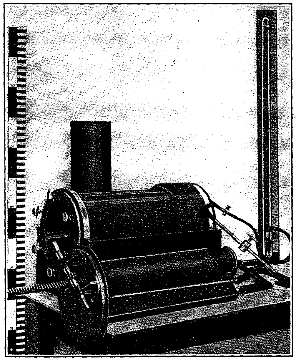
Pfaundler, in 1916, reviewed the previous methods of measuring body surface and elaborated a new method based on the principle that the body surfaces are usually in the form of a cylinder or obtuse cone. The body was divided into sixteen regions by use of an instrument- dermatograph, and the areas added to give the total surface. This instrument is illustrated in Fig. 13.
Respiratory Tract.-One of the most marked features of the premature and of the congenitally weak are the poor respiratory efforts, indeed, Billiard has defined congenital weakness as "the incomplete establishment of respiration." The premature in response to the need of air, inspires at birth, but its muscular power is weak and its efforts are insufficient to raise the thoracic wall and thus dilate the pulmonic cavity. As a result, though the large bronchi are filled with air, many of the small bronchioles are not dilated and a large portion of the lung continues to remain in a fetal stage, and may require several weeks for its complete expansion. The reason for this poor functioning of the organs of respiration lies in the lack of development of the respiratory centers in the medulla.
Most observers state that the chest wall of the premature infant is more or less immobile, moving but slightly with each respiration, but it has been our experience that quite constant evidence of prematurity is shown in the flexibility of the thorax and its tendency to retraction with each inspiration, the seeming immobility being the result of the poor effort on the part of the muscles of respiration, due to their weakness. The chest walls can expand but the muscular power is insufficient to make them do so. This muscular inertia, which is so well evidenced in these infants, is therefore partly the result of poorly developed muscles and partly the result of deficient innervation due to a similar lack of development of the cerebral centers.
Accompanying the deficient oxygenation of the blood are attacks of cyanosis, during which respiration ceases entirely. This apneic interval lasts for one or two minutes and then breathing is resumed. These attacks are not at all infrequent during the first fortnight and often appear without warning. In those cases in which recovery occurs the attacks become less frequent and less severe, but when unrelieved they are of grave significance and not uncommonly result fatally.
Clinically the weakened respirations. are manifested by the monotonous, feeble, whining cry and grunting expirations with comparative immobility of the thorax, and the superficial and often irregular character of the respirations, which become abdominal in type. While a child born at the sixth month may breathe for hours or days, previous to that time respiration is not fully established. Even though respiratory exchange does not occur, the heart may be found beating several hours after birth.
The frequency of respiration in the sleeping premature immediately after birth is frequently as high as 40 to 50 per minute. When awake the rate is about 50 or more unless the infant is crying, when it is much less than in ordinary breathing. The type of respiration in the premature is essentially diaphragmatic, superficial and irregular, showing interruptions particularly during crying when these pauses may be quite long. The soft and yielding character of the thoracic wall in the premature permits of slight degrees of retraction of the lower intercostal spaces during the deeper inspirations.
The physical findings over the lungs of premature infants are uncertain. On inspection and palpation the thorax shows deficient mobility, on percussion the, sounds over the bases are lower than over the balance of the chest, and on auscultation the vesicular murmur is hardly perceptible. At autopsy these signs are confirmed and the lower parts of the lungs particularly are seen to be atelectatic, at times the major portion of the organ being involved, making gaseous interchange very difficult.
The complete establishment of respiration may be prevented not only by the weakness of the respiratory movements but by the aspiration of liquor amnii or mucus during the last moments of delivery, which mechanically prevents the entrance of air into the pulmonary alveoli. (See Atelectasis.)
Parrot, Billiard and others have noted a condition which is spoken of as life without respiration, of which the characteristic manifestations are the absence of thoracic movements, the presence of a pulse and of movements of the extremities, and the absence of asphyxia immediately after birth. The persistence of the ductus arteriosus renders this condition supportable, as it allows the blood to pass directly into the aortic current without passing through the lungs. Such infants remain in their intra-uterine state of apnea until the respiratory centers become sufficiently irritated by the increasing venous blood to evoke respiratory action. This life without respiration should not be confounded with the apparent death of children born at or before term. Apparent death has two forms: The syncopal form, which is characterized by pallor of the skin and absence of pulse, and the asphyxiated form, distinguished by cyanosis of the skin and the presence of a pulse beat. (See Apparent Death.)
The nasal passages of the new-born prematures are particularly narrow, favoring the easy occurrence of stenosis in inflammatory conditions involving the nasal mucosa.
Interference with respiration also results from the aspiration of food or vomited matter into the larynx or trachea, the lack of development of the pharyngeal and laryngeal reflexes being responsible for the not infrequent occurrence of this accident. Attempts at drinking sometimes result in mechanical hindrance of obstruction to inspiration during the act of swallowing. Aspiration of food is often followed by a pulmonary infection and thus atelactasis of the lung may be said to predispose to a pneumonia which not infrequently leads to death. (See Infections of the Lungs.)
Jaschke considers the deficient function of the respiratory apparatus as being due to the fact that the irritability of the respiratory center is so low that a large accumulation of carbonic acid in the blood is necessary to make it act. With the sinking of the carbonic-acid tension with stronger respirations, the depth of respiration decreases again, because of lowered stimulation of the respiratory center and finally a point is reached in which the blood is arterialized, when the respiratory center no longer responds. A pause in respiration sets in and lasts until excess of carbonic acid stimulates new respiratory movements.
A further point is brought out by Jaschke. There appears to be a disturbance of the gaseous interchange, which is probably explained by the peculiarity of the blood serum of debilitated premature infants. This was first noted by Pfaundler. The blood serum shows a diminution of the OH ions, and a correspondingly greater concentration of the H ions, which condition makes the draining of carbonic acid from the tissues more difficult. Jaschke believes that this agrees with Finkelstein's theory that the attacks of cyanosis are to be regarded as an expression of a chronic carbonic-acid intoxication.
The Digestive Tract.-1. Anatomy.-The muscles of the buccal region, of the tongue and of the soft palate are weak.
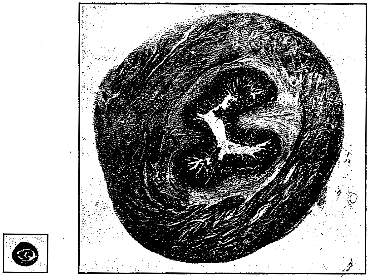
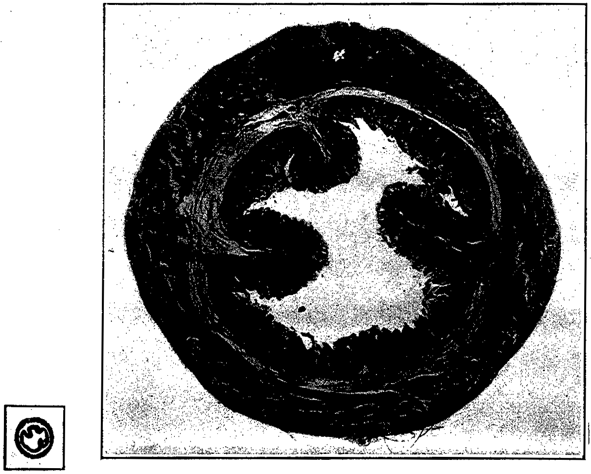
The stomach of the premature infant before its first feeding, as seen in autopsy, is in an almost vertical position and tubular in its form. In the premature infant which has been fed the fundus is fairly well developed and causes the stomach to assume a more oblique position. This is corroborated by a roentgen-ray examination (Figs. 14 and 15).
A. F. Hess was able to demonstrate that the gastric canal of the infant is more nearly vertical than horizontal, and that therefore from a functional standpoint the infant's food traverses the gastric canal in a vertical rather than a horizontal path, even though the stomach lies more or less horizontally. This fact is even more true of the physiological path of the food in the premature (Fig. 16).
The cardiac end of the stomach is found well to the left and usually about the level of the tenth dorsal vertebra. The cardiac sphincter is usually poorly developed (Fig. 17). This in part accounts for the ease with which the premature infant regurgitates its food. The pylorus lies somewhat higher than that of the full-term new-born, in whom it is found about midway between the ensiform cartilage and the umbilicus. Before feeding it is almost always found to the left of the median line. The pyloric musculature is usually quite well developed, even in the new-born premature (Figs. 18, 19 and 20).
The musculature of the stomach at autopsy in the new-born premature is in a state of contraction, giving the stomach a tubular appearance. In the living, however, this tubular appearance quickly disappears with the administration of food, the fundus enlarging much more rapidly than the balance of the stomach in order to meet the physiological demands.
Gastric Capacity.-Although many authors have measured the full-term infant's stomach as to its capacity, both at autopsy and in the living, their figures vary considerably.
Mosenthal, after a careful study of full-term infants measured during life and postmortem, states that the physiological capacity of the stomach exceeds the anatomical gastric capacity during life because of the rapid passage through the pylorus of the individual feedings during the act of nursing. This fact is corroborated by the roentgen ray (Fig. 16) in several of our cases. Therefore, the gastric capacity, as measured post mortem by filling the stomach with water under pressure of 15 cm. of water with the pyloric end of the stomach ligated, must also fall short of giving the exact functional capacity.
Pfaundler's figures for the stomach capacity during the first three months of life for the full-term infant are 90, 100 and 110 cc. Holt gives the following averages for stomach capacity in a series of studies made on infants dying during the first four weeks of life and examined postmortem.
|
Age |
No. of cases |
Capacity |
|
Birth |
5 |
36 cc. |
|
Two weeks |
7 |
45 cc. |
|
Four weeks |
4 |
60 cc. |
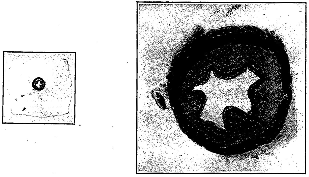
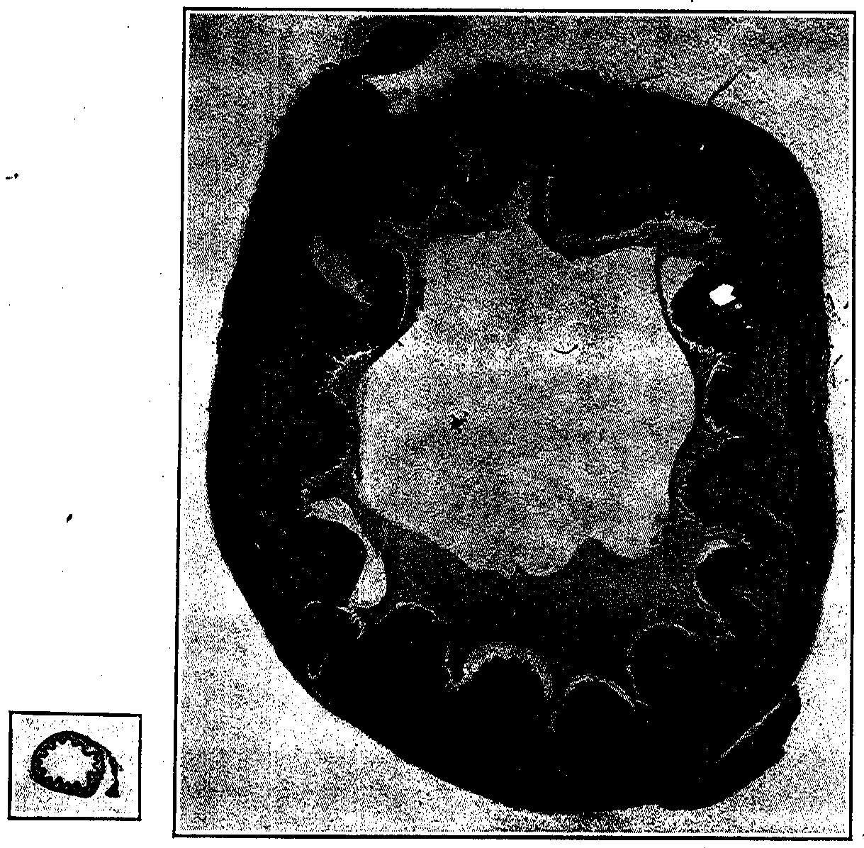
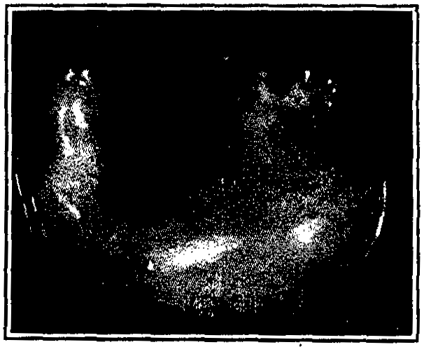
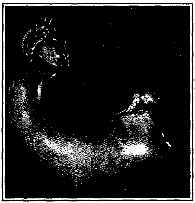
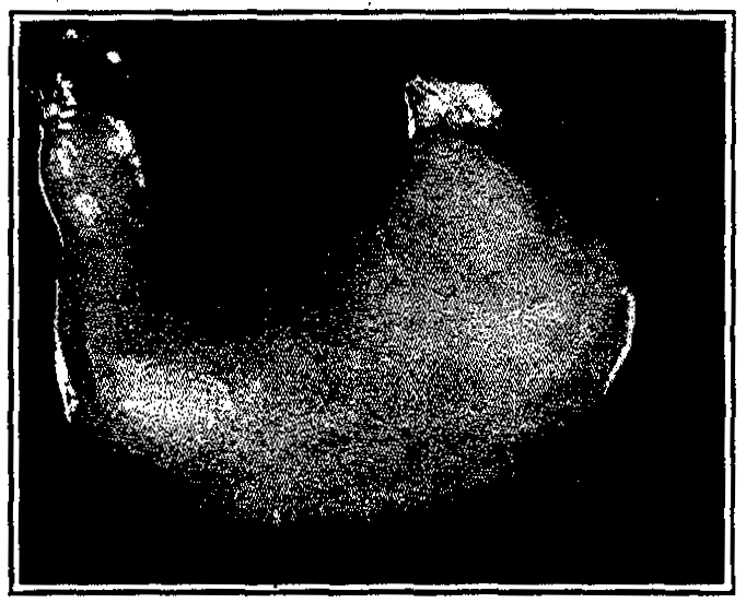
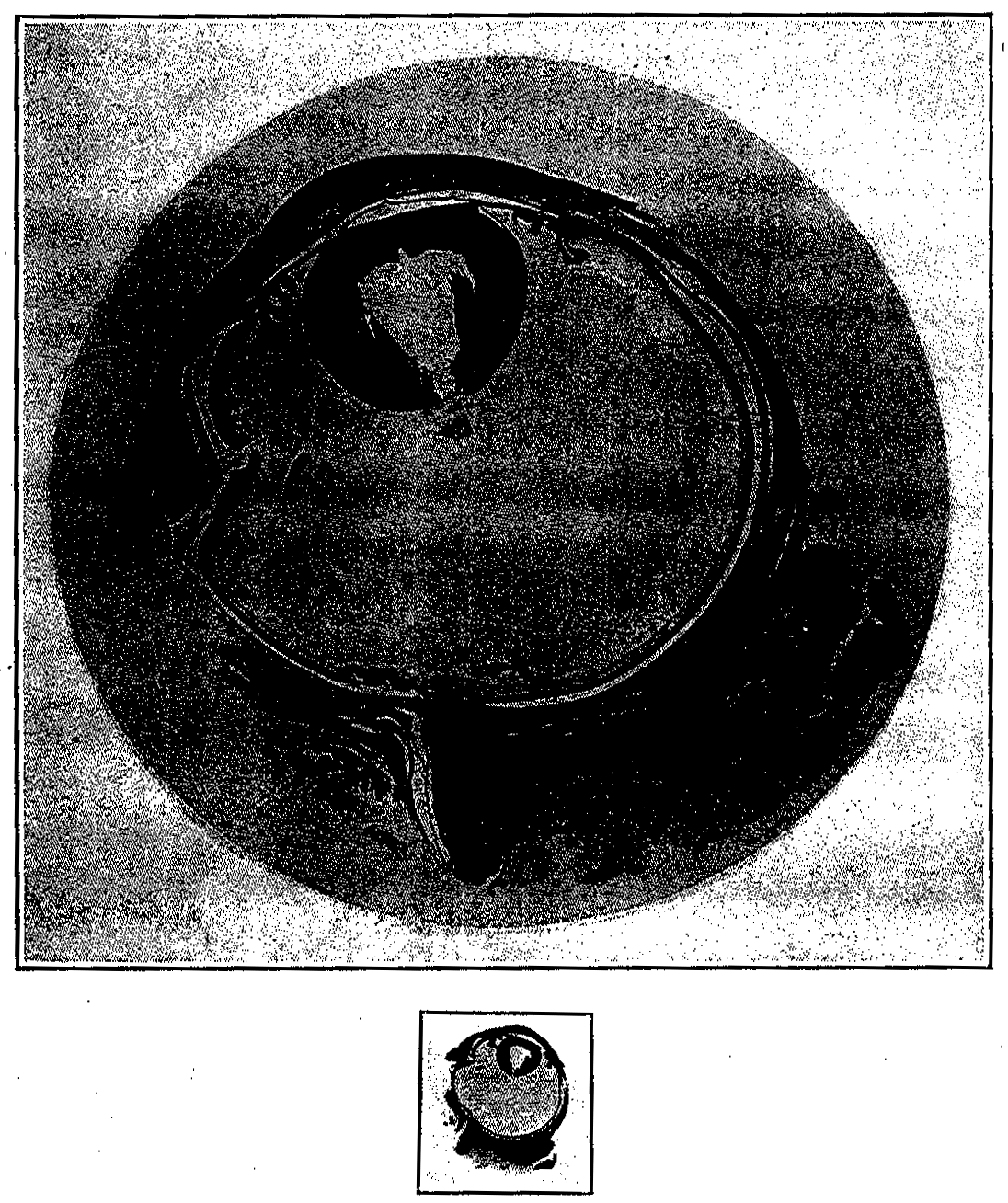
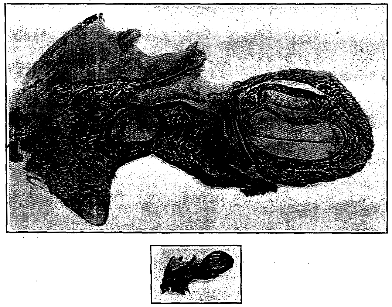
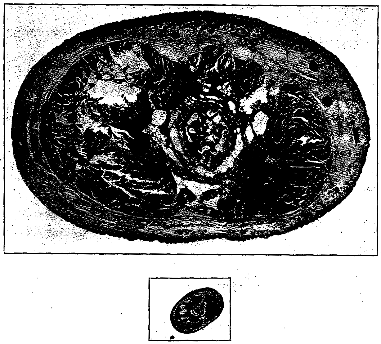
Notwithstanding the fact that distention of the stomach according to the method of Pfaundler at autopsy is far from an ideal method of estimating the physiological capacity of the stomach, the author has undertaken to measure the stomach capacity for the various fetal ages after the sixth month by this method, and to illustrate the same graphically by photographs which represent the actual size of these stomachs at various fetal ages. This has been done more especially to illustrate the dangers of individual overfeedings which are so disastrous to the life of the premature.
Figs. 21 to 26 are photographs taken with specimens immersed in oil and represent the exact size of the stomach under 15 cm. of water pressure at different ages.
The stomach of the premature infant on a diet of breast milk is usually found empty at the end of one and one-half to two hours. That of the artificially fed requires a considerably longer period of time, depending upon the nature of the food administered, even in the case of feeding with predigested milk.
2. Physiology.-The digestive functions of the healthy premature infant are proportionate to the age at the time of birth. At the sixth or seventh month most of the functions and secretions are rudimentary and insufficient, while in the older infants the lessening of digestive ability is not so great.
The sucking ability in the prematures and weaklings is feeble as a result of the lack of muscular strength necessary to operate the suction, the muscles of the buccal region, of the tongue and of the soft palate being weak. Accompanying this muscular asthenia is an inactivity of the salivary glands, as a result of which the mouth is dry. The lack of sucking movements tends also to retard the development of these glands.
The strength to swallow is also diminished in the premature. In the weakest a few drops of milk placed in the mouth remain there; in the stronger, though at first they nurse, they soon tire and their efforts to swallow cease.
"Hunger contractions" were studied by Taylor in 5 premature and 40 full-term new-born infants. A comparison of the contractions in the new born with several older children showed that the hunger contractions in the former were greater than in the latter. Reflex inhibition from the presence of food in the stomach was present in infants of all ages. The time of appearance of hunger after feeding in healthy infants gaining in weight and receiving a sufficient amount of food was: For premature infants under one month, one hour and forty minutes; in full-term infants under two weeks, two hours and fifty minutes; in infants from two weeks to four months, three hours and forty minutes.
The ferments of the gastro-intestinal canal are most conveniently discussed from the standpoint of action. The first group are those that aid in the splitting up of protein substances.
Pepsin is present in the gastric mucosa as early as the fourth fetal month, though not in such quantities as in the older children. It increases in amounts up to about the third month of life and then remains at about that level. Hydrochloric acid and rennin are also present in fetal life. Hess was able to demonstrate free hydrochloric acid in 54 out of 55 cases immediately after birth.
Lipase was found to be present in small quantities by Ibrahim in a fetus of 800 gm. and plainly present in those from 1100 gm. upward Sedgwick had previously demonstrated it in 1906.
Trypsin is present in the pancreatic extract of the new born. Ibrahim found trypsinogen as early as the sixth fetal month and enterokinase was also found by him in an extract of intestinal mucosa from premature infants. The lower third of the small intestine is most active in the production of enterokinase.
Secretin, the ferment which activates the pancreas, was found in the small intestine of the full-term new born by Ibrahim and Gross, but its activity was slight. In the premature it is probably even more deficient.
Erepsin splits albumoses and peptones and originates from the mucosa of the small intestine. It has been demonstrated in the premature by Langstein, Jaeggis, Cohnheim and others.
The next group consists of the carbohydrate ferments, of which the milk-sugar ferment lactase is found in the intestinal contents, the stools and the intestinal mucosa. It is frequently absent from the intestinal tract of the premature, as it makes its appearance rather late in fetal life. Nothmann was able to demonstrate it in the stools of the mature new born in only a few cases. The presence of relatively large amounts of milk sugar in the infant's . food probably increases the amount and activity of the lactase. The deficiency of lactase at birth is indicated further by the finding of lactose in the urine of new-born infants (Nothmann). This would seem to point to a lack of milk-sugar fermentation (von Reuss).
The cane-sugar splitting ferments, invertin and saccharase, are present at an early date in embryonal life, although there is no use for them in those fed on human milk or where lactose is used artificially, for a long period of time. They are found in the intestinal walls and in the meconium.
Maltase is present, according to Ibrahim, in all parts of the small intestines and in the intestinal contents of prematures. Diastase, the amylolytic ferment, is present in the salivary glands and in the pancreas of the new born. Ptyalin is found in the parotid and in the submaxillary secretions, although it is not required until the beginning of the starch feeding. Ibrahim believes that the pancreatic function of the new born and especially of the premature new born is somewhat below that of the older infant, and, therefore, the instructions of the older clinicians not to feed these infants mixtures containing much starch were correct from a physiological point of view (von Reuss).
The third and last group of. ferments are those which act upon the fats. Steapsin was found in the pancreatic secretion by Zweifel, and Ibrahim showed that it was also present in the premature. The meconium contains this ferment. Lipase is very active in the gastric mucosa of prematures.
In general the premature may be said to possess nearly all the ferments necessary for the breaking-down of its food. Some of them, such as diastase and ptyalin, which are not. present during fetal life or only in the most insignificant quantities, are called forth even in the premature, by the administration of food, and though they may be deficient both in amount and in activity at this time, the continued stimulation offered by food soon results in a material increase in both qualities, at least in the case of prematures who possess a sufficient degree of vitality. All necessary ferments being present, it is of little advantage to feed the premature infant predigested human milk.
Ferment therapy also is of little value in premature infants as is also true in older children. If the required ferments are present they will increase with the giving -of food. It is not the absence of ferments that is responsible for the peculiarities of action of the digestion of the prematures, but rather the way the food is broken down and absorbed; and a clear realization of these differences is necessary to an understanding of the peculiarities of the digestion of the new born, both premature and at term.
The normal gastric mucosa provides only for the absorption of salts and carbohydrates.
Ganghofer and Langer' found that up to the fourth day of life the intestinal tract is permeable to foreign proteins and the importance of this is great. The permeation of. these through the intestinal wall results in the formation of antibodies in the tissues, and the danger of sensitization of the organism to that particular protein. Herein lies one of our most important indications for feeding with human milk.
The intestinal canal is more frail than in the full-term infants and the intestinal musculature is weak and easily distended and often times unable to expel the contained meconium.
The meconium begins to be formed at the fourth fetal month. It is made up of the secretions of the gastro-intestinal tract, vernix caseosa, threads of mucus, desquamated epithelium, biliary acids and salts, cholesterol, fat droplets, fatty-acid crystals and liquor amnii which has been swallowed. That which is passed on the first day is dark green; thick, sticky, homogeneous and odorless. Its excretion lasts from twenty-four to ninety-six hours. During the first few hours it is free from bacteria and even later the number of organisms present is small. The characteristic yellow color of the breast milk stool is scarcely established before the fifth and sixth day and then only when the milk taken is rich. The sour odor of the breast-milk stool may also be recognized at this time.
Hymanson and Kahn, investigating the properties of meconium found that there were traces of ammonia and amylase, but no uric acid, trypsin, erepsin, lactase or lipase. Their analysis of the inorganic constituents is given in the table which follows:
|
|
1 |
2 |
3 |
4 |
5 |
|
Parts per thousand: |
|
|
|
|
|
|
Water |
732.3 |
801.7 |
784.5 |
697.7 |
718.6 |
|
Dry matter |
267.7 |
198.3 |
215.5 |
302.3 |
281.4 |
|
Organic matter |
245.2 |
180.1 |
197.7 |
280.5 |
257.9 |
|
Ash |
22.5 |
18.2 |
17.8 |
21.8 |
23.5 |
|
Ash percentage of: |
|
|
|
|
|
|
Total meconium |
2.25 |
1.82 |
1.78 |
2.18 |
2.35 |
|
Dry matter |
8.3 |
9.1 |
8.2 |
7.2 |
8.3 |
|
Analysis of meconium ash (per cent) |
|
|
|
|
|
|
Fe2O2 |
3.17 |
1.17 |
2.24 |
0.92 |
1.44 |
|
CaO |
18.24 |
|
17.55 |
21.18 |
16.34 |
|
MgO |
4.21 |
8.05 |
6.17 |
6.18 |
4.75 |
|
P2O5 |
12.62 |
8.62 |
|
|
11.70 |
|
SO2 |
23.14 |
25.63 |
18.47 |
28.30 |
24.32 |
|
CI |
5.86 |
7.12 |
6.89 |
5.34 |
|
|
K and Na |
24.19 |
|
3.72 |
|
|
3. Bacteriology of the Gastro-intestinal Tract.-The gastro-intestinal tract of a healthy premature is sterile at birth and remains so for a short time afterward, the meconium remaining sterile for about twelve hours. This is followed by invasion of bacteria, most probably with the first feeding, and during the next two days the gastro-intestinal flora is very variable, depending chiefly on the surroundings of the infants. After the third day, however, a typical intestinal flora develops, the type depending chiefly upon the diet of the infant.
In an infant fed with human milk saccharolytic bacteria predominate, the chief one being Bacillus bifidus, which is especially numerous in the large intestine up to the sigmoid flexure. This portion of intestine also contains the largest number of bacteria. Bacillus coli is also present, especially in the region of the ileocecal valve and cecum, but still Bacillus bifidus predominates. The flora of an infant on human milk are much more homogeneous than that of an infant artificially fed.
In artificially-fed infants there is a relative increase of Bacillus coli and of proteolytic bacteria and a diminution of Bacillus bifidus. However, the flora of artificially-fed infants are much more variable and depend chiefly on the chemical composition of the food.
Human milk low in protein and high in sugar leads to the flora of fermentation, while cows' milk which is high in protein and low in sugar leads to flora of putrefaction.
Carbohydrates favor the development of fermentative organisms, lactose favoring especially Bacillus bifidus and maltose and dextrin compounds favoring Bacillus acidophilus.
Proteins favor the development of organisms of putrefaction, especially when given in excess.
Fat seems to have no distinctive action on the intestinal flora.
Metabolism of Premature Infants.-The following facts are quoted from Jaschke, who states that there is not sufficient material on hand at the present time for comparing the metabolism of prematures both healthy and debilitated with the metabolism of mature normal infants.
"The expenditure of energy as related to the unit of body surface is in the premature much greater than in the mature new born (Camerer), when the age is calculated from birth; on the other hand, however, they are almost the same, if age is calculated from the time of conception (Pfaundler) which very well agrees with the curve of the potential of life. The nitrogen under balance in the premature lasts longer than in the mature new born, which is probably dependent in the first place upon the small food intake. There are not sufficient experiments on the gaseous exchange and on the insensible perspiration to enable one to draw conclusions that would be of general value. There is nothing known of mineral metabolism."
Nervous System.-The lack of development of the cerebrospinal nervous system is greater than that of the sympathetic system. It is most markedly evidenced by the muscular inertia shown by the infant. Many of them lie in a state of stupor or somnolence from which they must be aroused to be fed. Others can be aroused by external stimulation which calls forth only a weak cry and slight movements of the body. These movements are slower than those of the full-term infant and the child tends to relapse into a deep sleep as soon as the stimulus is removed. Also depending to some extent upon the incomplete development of the nervous centers are the weak respiratory functions and the feeble efforts at sucking. A this time the development of the brain is still going on and the separation of the white and gray matter is not yet completed.
The nasal and pharyngeal reflexes are particularly weak in children born before term. Abdominal reflexes are almost never present in the premature; in fact they are rarely seen in the new-born infant.
Among many neurologists the opinion is prevalent that prematurity predisposes to idiocy, imbecility and epilepsy. However; it appears in these instances it is not so much the premature birth that is responsible, but rather there seems to be a common cause leading to retarded development and premature expulsion of the fetus.
Cardiovascular System.-As compared with other organs the heart is relatively well developed. That the heart should be strong is not surprising, as from the first months of pregnancy the precocious development of this organ is found to be in complete accord with the importance of its function. The high position of the diaphragm and the equality of the diameters of the thorax causes the long axis of the heart to lie in a more nearly transverse position. Because of this position the apex beat is found in the fourth interspace, 0.5 to 1 cm. outside the mammillary line.
The variability of the pulse-rate, which is quite marked in the premature new born, ranges from' 90 to 200 per minute, with an average of about 120. This variability is the result of the lack of development of the cardiac inhibitory centers.
At birth the thoracic respirations determine a considerable flow of blood through the pulmonary artery to the lungs. The function of the ductus arteriosus ceases at this time and the blood current is diverted from the foramen ovale through the tricuspid orifice into the right ventricle. Within twenty-four to forty-eight hours after birth the ductus arteriosus is almost completely closed normally, while the foramen ovale is soon completely occluded by the rapid growth of its valvule. If, however, the ductus arteriosus is not closed, as is frequently the case in the premature infants, due to non-expansion of the lungs with a resulting increased resistance in the lesser circulation, cyanosis may result.
The heart is usually only secondarily involved in asphyxial attacks, the tones becoming weak and slow during the spells of cyanosis: The heart action often persists for hours after the respiration ceases. Myocardial asthenia in the premature may also result in cyanosis and is frequently accompanied by edema. (See Cyanosis.) General circulatory difficulties may also be the cause of subnormal temperature in these infants.
Blood-pressure in the mature at birth and for the first few days of life is low and in the prematures and weaklings it is still lower. In the new born the pressure ranges around 80, while the figures for the premature and the weaklings will vary from 60 to 70 mm. of mercury (Trumpp).
The vascular walls in the premature are weaker than in the infant at term and because of this these children are subject to hemorrhage following relatively slight traumata. This is particularly true of the intracranial vessels and thus we see that hemorrhages in this region are relatively more frequent in the premature.
The intracranial hemorrhages are usually followed by early death and in many instances undoubtedly these are interpreted as respiratory deaths because of the influence of pressure on the respiratory center.
This tendency to hemorrhage in the premature in some cases is due to deficient coagulability of the blood.
In a study of the new-born Rodda found by his method that the average coagulation time was seven minutes, with a normal range between five and nine minutes.
There is a prolongation of coagulation and bleeding times from the first day to the maximum on the fifth day of life, with a return to the average first-day determination time before the tenth day. It is significant that this coincides with the age incidence of hemorrhagic disease and cerebral hemorrhage.
In icterus neonatorum normal coagulation and bleeding times were found.
Several cases of melena neonatorum gave markedly prolonged coagulation times-up to ninety minutes-and bleeding times of hours, days. or until the condition was controlled.
Suspected and mild cases of congenital syphilis gave normal findings. Severe and progressive cases gave prolonged times. Pfaundler found a low alkali reserve in debilitated prematures and believed this to be an important factor in the low immunity to infection. The blood also shows an increased viscosity due in all probability to the increase of water loss over intake during the first days. Rusz has also suggested, as a second factor, the delayed tying of the cord, with a resulting flow of blood from the placenta, causing a relative overloading of the fetal circulation.
The cell content of the blood of the premature does not differ greatly from that of the new-born infant, though it does possess certain special characteristics (Kunckel).
The erythrocytes are slightly less in number and diminish readily under the influence of infections, jaundice and edema. Macrocytes and microcytes are very numerous and poikilocytosis is also often observed. Nucleated erythrocytes are characteristic of the blood of the premature infant and the farther the child is from term the more numerous are these nucleated cells. In the mature infant nucleated cells are only found during the first few days of life, while in the premature they are found as late as ten days after birth. A large number of these cells is incompatible with life. They re-appear quickly with the onset of any infection and are slower to disappear when the temperature is subnormal. With redevelopment of a subnormal body temperature during the first weeks of life, the nucleated reds tend to reappear.
The leucocytes are less numerous or only slightly increased. Instead of 12,000 to 13,000 leucocytes, as found in the normal full-term new-born infant, there are on the average 8000 in one cubic millimeter in the premature.
The differential count shows a high percentage of mononuclears and abnormal elements, such as mast cells (basophiles) and myelocytes. These cells possess little activity, which is an important factor in the low resistance of these infants to disease. What bearing the lowered alkalinity has on the feeble reaction of the white cells is still an open question. The reaction to infection is, as a rule, very feebly polynuclear and may even be replaced by one of transitional forms and abnormal elements, myelocytes and mast cells, as if the hematopoietic organs were only capable, in their deficiently developed states; of putting into the circulation immature elements. The polynuclear eosinophiles are fewer in number in the premature and disappear when an infection occurs. In congenital syphilis they are usually increased.
While in the normal full-term infants the hemoglobin content gradually sinks and at the end of the fourth week amounts to about 85 per cent (by Sahli's hemoglobinometer), in the prematurely born infants its value at this time is 50 to 60 per cent; therefore, in prematurely born infants there is a distinct and very early hemoglobin impoverishment of the blood, which reaches its maximum in about the third to the fourth month. While the hemoglobin content shows a marked deviation from the normal, the number of erythrocytes is only little below the normal and therefore the hemoglobin content of the individual blood corpuscle is considerably less than normal. This accounts for the constant and early development of anemia in prematures during the first three months of life. The cause of this hemoglobin deficiency seems to be an insufficient iron content of the premature's blood, which is easily understood when we recall that Hugouneng has proven that the quantity of iron stored up by the fetus in the last third of pregnancy is twice as large as that during the first two-thirds.
Lichtenstein's studies on a large number of premature infants showed a considerable degree of anemia in a large percentage of his cases.
In a study of the blood findings in 90 prematurely born infants (those of known syphilitic and tuberculous parentage were excluded) in greater part born one or two months before term, Lichtenstein recorded the following findings:
In 10 cases he found:
|
Age |
Hemoglobin |
Red blood cells |
White blood cells |
|
First day of life |
96.7 |
6,135,000 |
7,512 |
|
Third day of life |
90.7 |
5,799,000 |
5,755 |
|
Tenth to twelfth day of life |
85.8 |
5,376,000 |
8,572 |
The hemoglobin and red cell counts were relatively those of the full term, showing an absence of congenital enamia. The white blood cells were below the averages given for full-term infant and presented an absolute leukopenia.
There was also a more marked anisocytosis, polychromatosis and erythroblastosis than is seen in the blood picture of the full-term new-born.
Subsequent examinations of the blood in 19 premature infants breast fed over two weeks by healthy mothers gave the following averages:
|
Age |
Hemoglobin |
Red blood cells |
White blood cells |
|
3 to 4 weeks |
76.0 |
4,023,000 |
7,560 |
|
2 months |
50.5 |
3,616,000 |
8,720 |
|
3 months |
40.2 |
2,945,000 |
7,042 |
|
4 months |
40.5 |
3,065,000 |
7,969 |
|
5 months |
44.0 |
3,733,000 |
7,969 |
|
6 months |
40.0 |
3,740,000 |
7,969 |
These results, when compared with examinations of wet-nurses' infants, showed a decided oligochromemia (controls never under 55); and oligocythemia (controls rarely under 4,000,000). The red cells increased toward the end of the first half year of life. The white cell counts for full-term infants usually averaged between 10,000 and 12,000, those of the prematures after the fourth week between 7000 and 9000.
There is also a constant anisocytosis and anisochromemia which changes run parallel with the oligochromemia. Erythroblasts were found in some cases as late as the fourth month.
The percentages of the various white cells do not vary greatly from the picture of the full-term infant. Metamyelocytes were occasionally seen as late as the second month. The figures are tabulated in the following tables:
|
Day of examination |
Neutrophile leucocytes, per cent |
Eosinophile leucocytes, per cent |
Small lymphocytes, per cent |
Other lymphocytes, per cent |
Large mononuclears, per cent |
Metamyelocytes, per cent |
|
1st day |
45.8 |
1.8 |
11.6 |
18.5 |
8.2 |
13.4 |
|
3rd day |
31.0 |
3.1 |
23.5 |
27.5 |
8.7 |
5.9 |
|
10 to 12 days |
27.9 |
3.2 |
20.3 |
33.2 |
11.2 |
3.8 |
WHITE BLOOD CELL PERCENTAGES IN THE LATER MONTHS (LICHTENSTEIN)
|
|
|
|
|
Eosinophile |
||||||
|
Age of infants |
Max per cent |
Min per cent |
No. cases |
Max per cent |
Min per cent |
No. cases |
Max per cent |
Min per cent |
No. cases |
Per cent |
|
0.5-1 month |
33.5 |
5.8 |
5 |
76.5 |
51.5 |
5 |
14.5 |
1.0 |
5 |
2-3 |
|
2 months |
20.3 |
7.3 |
8 |
82.8 |
56.5 |
8 |
17.3 |
6.3 |
8 |
2-3 |
|
3 months |
41.5 |
9.5 |
6 |
79.8 |
44.0 |
6 |
12.0 |
3.5 |
6 |
2-3 |
|
4-6 months |
35.0 |
29.8 |
4 |
60.8 |
48.0 |
4 |
14.3 |
4.8 |
4 |
2-3 |
Lande examined a group of 70 prematures born from the. sixth to the eighth month of pregnancy, with weight from 830 to 2500 gm. The majority were fed on human milk, made an average monthly gain of 450 gm., and were relatively free from infection and congenital lues. The results of examination of the hemoglobin content and the percentage of the red and white blood cell elements in the newly born prematures during the first weeks of life are shown in the following table:
|
|
|
|
|
|
|
|||||||
|
Age of infants |
No. cases |
Max per cent |
Min per cent |
Commonest value, per cent |
Max cc |
Min cc |
Commonest value |
Max |
Min |
Max |
Min |
Commonest vlaue |
|
1 day |
12 |
140 |
100 |
110-120 |
5.8 |
3.8 |
4.3-5.0 |
7000 |
400 |
20,000 |
3800 |
10,000 to 15,000 |
|
2-4 days |
15 |
135 |
100 |
125 |
6.7 |
4.1 |
4.6-5.4 |
6700 |
0 |
16,000 |
3600 |
8,000 to 12,000 |
|
6-8 days |
6 |
105 |
100 |
|
5.6 |
4.0 |
|
160 |
0 |
11,400 |
6600 |
|
|
|
|
|
|
|
|
|
|
||||||
|
Age of infants |
No of cases |
Max per cent |
Min per cent |
Commonest valu e per cent |
Max per cent |
Min per cent |
Commonest value per cent |
Max per cent |
Min per cent |
Commonest value per cent |
Eosinophiles |
Mast cells |
Myeloblasts and myelocytes |
|
1 day |
12 |
54 |
12 |
40-50 |
85 |
39 |
45-55 |
12.5 |
1 |
7-10 |
0.5-1.5 |
0-1.5 |
3-12.5 |
|
2-4 days |
15 |
64 |
11 |
40-55 |
87 |
30 |
40-55 |
12.0 |
2 |
5-10 |
0-5.0 |
0-2.5 |
2-6.0 |
|
6-8 days |
6 |
68 |
29 |
|
65 |
26 |
|
8.0 |
6 |
|
0-2.0 |
0-2.0 |
0-2.0 |
From these tabulations Lande drew these conclusions: Opposed to the findings in full-term infants; there is in prematures a greater richness of nucleated red blood corpuscles, a more frequent appearance of myeloblasts and myelocytes during the first days of life, a lesser development of absolute and relative leukocytosis, and a greater number of immature leukocyte forms.
The blood picture from the third week of life to the age of six months is expressed by the following figures (Lande):
|
|
|
|
|
||||
|
Age of infants |
No. of cases |
Max per cent |
Min per cent |
Commonest value per cent |
Max, Millions |
Min, Millions |
Commonest value, Millions |
|
1.0 month |
13 |
105 |
70 |
80-85 |
5.5 |
33 |
3.6-4.6 |
|
1.5 month |
9 |
95 |
50 |
60-70 |
3.9 |
2.7 |
3.2 |
|
2.0 month |
17 |
80 |
50 |
60-70 |
4.4 |
2.4 |
3.0-3.6 |
|
2.5 month |
7 |
80 |
50 |
60-65 |
4.0 |
2.3 |
3.0-3.6 |
|
3-3.5 month |
24 |
80 |
50 |
60-70 |
4.9 |
2.4 |
2.9-3.9 |
|
4-4.5 month |
18 |
75 |
50 |
60-70 |
5.2 |
2.7 |
3.3-4.0 |
|
5-5.5 month |
15 |
85 |
55 |
65-75 |
4.7 |
3.4 |
3.9-4.6 |
Lande noted a fall in the hemoglobin content from 80 to 85 per cent to 60 to 65 per cent in the third month, which slowly rises to 65 to 75 per cent in the sixth month. At the same time the number of erythrocytes sinks from about 4,000,000 to 3,300,000, in order to again approach the normal value by the end of the first year.
Nathan and Langstein have found the blood in the new-born very low in antitoxic, bactericidal and hemolytic properties.
Blood-sugar determinations in three healthy prematures fed on mother's milk were studied by Heller. In no case was sugar found in the urine, this being explained by the fact that in no instance was there an evident hyperglycemia. The percentage of blood sugar noted between ten and a half and twelve hours after birth was 0.095, 0.089, 0.082; these figures are for infants weighing respectively 1500 gm., 1380 gm. and 930 gm. The diets were increased so that on the seventh day the two heavier infants were both getting 160 gm. while the smaller was given 80 gm. of milk. The percentage of blood sugar was then noted; 0.104 for both heavy infants (twins) and 0.065 for the other. All observations were taken from one-half to two hours after the feeding. There was a steady fall in blood sugar in the twins until the sixth day.
These blood-sugar findings are similar to those of Gotzky, who found an average of 0.085 mg in the full-term new born, somewhat lower findings in prematures, and 0.095 mg in later infancy, as compared with 0.102 mg in later years. Because of the relationship of blood sugar to diet, comparative studies must be undertaken with a knowledge of the quantity and quality of food and the time to the meal.
Lymphatic System.-This is well developed and does not differ materially from that of the new born, unless possibly its circulation is slowed as a result of the slowing of the general circulation.
Thymus and Thyroid Glands.-These organs present the highest degree of development of any glandular structures. In fetal life these organs contribute to the formation of blood and during the first few weeks of life have a phagocytic action.
Genito-urinary System.-In the female the labia minora usually overlap the labia majora, while in the male the testicles are often high in the inguinal canal, though it can happen that they are found in the scrotum as early as the seventh month.
An examination of the urine of the premature throws but little light upon the metabolism of these infants. The proportion of ammonia N to the total N is below normal, while C/N is increased. This speaks for an increase in the processes of decomposition. Nobecourt and Lemaire found that the freezing-point of the urine of prematures was lowered.
The amount and character of the urine during the first few days of the life of the premature depend upon the intake of fluid, upon the absolute body weight and upon the absolute and relative amounts of water within the body tissues.
If the quantity of fluids taken is small the amount of urine secreted is proportionately small. When the quantity is larger, as is usually the case if the infant is given feedings to substitute the mother's milk and frequent feedings of water, the relative as well as the absolute amount of urine secreted is larger. Cramer found that with an abundant supply of milk during the first few days of life the urinary output amounted to 54 to 60 cc for every 100 cc of milk consumed.
The frequency of urination during the early days of the premature is less than at an older age. While the infant may urinate during its passage through the birth canal or immediately after, yet, as a rule, during the first few days the urinations are very infrequent, at most three or four and more often only one or two times during the twenty-four-hour period. It is not at all uncommon that no urine is passed during the first day. This failure to urinate during the first day of life is not of much moment, but in those instances of absence of urination for as long as four days, as have been reported, some anomaly was undoubtedly present. With the increase in the fluids taken, which occurs usually on the third or fourth day, the frequency of urination also increases.
During the period of greatest concentration the reaction of the urine is strongly acid. As it becomes more dilute, the acidity becomes less marked.
Albuminuria is a symptom shown by almost all infants just after birth, full-term as well as premature. The length of time during which this persists is short, seldom more than the first few days, and the quantities of albumin present are small: 0.25 gm. of albumin per 100 cc of urine is a maximum which is frequently reached in the full-term infant. Von Reuss found the urine of only 4 per cent of new-born infants to be free from albumin during the first four days of life. After that time the amount of albumin present rapidly falls, unless the concentration of the urine remains high, and it retains the turbidity characteristic of infant urine, when the albumin persists for a longer period.
Albumin in the urine of the new born would seem to be somewhat of a physiological condition, certainly having no relation of a causal nature to the infections or other toxic factors of the later periods of life. Albuminuria at this time seems to have a certain analogy with the orthostatic albuminuria of older individuals, both probably to be accounted for by circulatory disturbances of the kidney. Von Reuss believes the condition is most easily explainable on the basis of circulatory stagnation which occurs in a more or less pronounced degree after every delivery. Uric-acid infarcts may also have some bearing as a cause of albuminuria. The deficient blood supply of the kidney and the lack of water passing through the organ as a result of the small quantity taken during the first few days of life probably increases the amount of albumin passed.
Nothmann found milk sugar in the urine of premature infants who were breast fed, and he reports that he found no such cases among the full-term infants. Sugar was found by Hoeniger in the urine of several infants delivered by forceps. It was excreted for three or four days and then gradually disappeared. It was believed to be the result of the force used during the operative delivery.
Acetone bodies are found in small quantities in the urine of poorly nourished and underfed weaklings.
During icterus neonatorum bilirubin occurs in the urine in the form of a precipitate. It is also found in solution in septic conditions and .in hemorrhage of the new born.
Creatin and creatinin have not been studied in the premature. Occasionally hyaline casts in small numbers, often covered with urates, are seen. These are probably due to the same causes as the albumin and have no pathological significance.
Special Senses.-Over the eyes of the youngest prematures occasionally there can be seen more or less well-marked vestiges of the pupillary membrane, the cornea is inclined to be somewhat thicker, the. anterior chamber less deep and the iris less pigmented. Strong light impressions are followed by reflex closure of the lids, but sudden movements are not followed by such closure, as the reflex is psychic, depending upon fear.
The eye movements of the premature infant are incoordinated, motion being most often in a horizontal direction, occasionally outward, but more often and in a comparatively strong manner, inward. It is not uncommon to see this tendency to convergence persist until the second month. The light reflex is present before birth and the pupil, when exposed to a strong light, contracts only to dilate again in two or three seconds. This secondary dilatation is particularly well marked in the premature as a result of the poor development of the nerve fibers, which are easily fatigued (Furmann). The convergence reflex is absent in prematures as well as in the more mature infant because fixation does not occur.
Skin and Adnexa.-The skin is thin, soft and usually of a more or less vivid red appearance, occasionally of a peculiar cyanotic hue, and the transparent dermis allows the circulatory network to be clearly distinguished. The skin is partly or completely covered with lanugo hairs which are seen most commonly between the shoulder blades, but also frequently upon the face and upon the extensor surfaces of the extremities. There is also noted extensive milium and flaccidity of the auricle and alae nasi, whose cartilage is not properly developed.
Icterus is usually more pronounced than at term and erythema is slower to disappear. If hypothermia develops the redness of the skin usually fades.
The absence of subcutaneous fat betrays itself by an angular appearance of the face, the chin is pointed, the head is small and narrow and the wrinkles of the skin impart an oldish appearance to the face which is especially marked after a few days when the loss of weight has been material and the skin often hangs in folds over the muscles and bones. Not infrequently the skin appears glossy as if on tension and this is seen especially in small prematures in the presence of sclerema and scleredema. Patches of skin may be absent, especially over the heels.
The hairs on the scalp are short and feebly colored, the nails are often poorly developed and do not reach the end of the fingers or toes and the rose is covered with small white comedones. The navel is closer to the symphysis than at term.
Mammary Glands.-The mammary glands are, as a rule, poorly developed, usually not palpable and particularly in the younger prematures do not often attempt to secrete milk. If fluid is present, as it may be in the older prematures, it usually makes its appearance about the eighth day, is most abundant up to the fifteenth day and may last until the third month. It is equally common in either sex. In most cases the secretion amounts to only a few drops, but occasionally larger quantities are seen.
Skeletal Development.-The lack of exact anatomical data as to the skeletal development of the premature infant has caused the author to resort to the use of roentgenographic studies. The stage of ossification of the skeleton of the fetus as observed in roentgenograms is of considerable practical importance in determining the age of the fetus. In addition observation by the roentgenographic method is more reliable than determination of age based on length and other measurements, since osseous development is more regular and offers many more factors for consideration. Pathology may often be readily recognized. Our studies thus far have shown that in the early months more accurate determination is possible than in the later months, because many more new centers appear in the first months, and the time of appearance is more constant.
The study of the roentgenograms for diagnostic purposes discloses that the cephalad segments, including the upper axial skeleton and upper extremities, are far more constant as to time of development of their osseous centers than the caudad segments and those of the lower extremities. This should be borne in mind in making comparative studies.
The figures as to length and other measurements of the fetus have, been discussed earlier (p. 29). Basing our facts on the roentgengraphic studies of a series of 55 normal cases, whose ages were determined from the history of menstruation and pregnancy and from their measurements, the normal process of development of the human skeleton was found to be as follows (Fig. 29).
Other Measurements of the Fetus.-Von Winckel regards the circumference of the head as of importance for the diagnosis of the age of the fetus and gives the following figures:
|
4th month |
10-14 cm |
|
5th month |
13-18 cm |
|
6th month |
19-24 cm |
|
7th month |
23-28 cm |
|
8th month |
25-30 cm |
|
9th month |
29-33 cm |
|
10th month |
32-37 cm |
The weight is entirely unreliable for the estimation of the age of the fetus, because it is subject to too many variations and is much influenced by the mother's general condition, and more especially by her diet.
Thus, it is seen that even the length, which up to this time has been regarded as the most reliable criterion for the determination of the age of the fetus, has many shortcomings and may result in an error of several weeks.
The ossification of the human skeleton begins in the upper part of the body and spreads very rapidly in both directions.
Seventh Week.-The first centers of ossification develop in the clavicles in the sixth to seventh week of intra-uterine life (Kreibel-Mall, Rauber-Kopsch), but they do not become visible in the roentgenograms until in the seven week. The ossification center appears in the middle of each clavicle and spreads rapidly in both directions.
Soon after the ossification has started in the clavicle one center appears in each half of the mandible.
Outside of these centers of ossification usually no other centers, except occasionally that of the maxilla, are visible in roentgenograms of the seven weeks' old fetus.
Eighth Week.-Osseous development makes rapid progress in the eighth week, and a large number of centers of ossification become visible at this time.
The following bones show centers of ossification demonstrable in roentgenograms.
Skeleton of the head: The squamous portion of the occipital bone and superior maxilla. In the latter the ossification begins soon after that of the mandible, the center appearing above. the region where the alveolus of the incisor tooth is later located.
|
Mandible |
7th week |
|
Occipital bone (squamous portion) |
8th week |
|
(lateral and basilar portion) |
9th to 10th week |
|
Superior maxilla |
8th week |
|
Temporal bone (petrous, mastoid and zygoma) |
9th week |
|
Sphenoid (inner lamella of pterygoid process) |
9th week |
|
Sphenoid (great wings) |
10th week |
|
Sphenoid (lesser weeks) |
13th week |
|
Sphenoid (anterior body) |
13th to 14th week |
|
Nasal bone |
10th week |
|
Frontal bone |
9th to 10th week |
|
Bony labyrinth |
17th to 20th week |
|
Milk teeth (rudimenta) |
17th to 28th week |
|
Hyoid bone (greater cornua) |
29th to 32nd week |
Usually no centers of ossification are present in the axial skeleton in this week.
Shoulder girdle: In the scapula a center of ossification usually appears in the eighth. week, sometimes in the ninth week. The center corresponds to the position of the middle of the spine of the scapula.
Upper extremity: The humerus is the first bone of the free extremities to show a center of ossification, which appears in the diaphysis early in the eighth week. Radius and ulna follow in the order given, the centers appearing very soon after those of the humerus.
The ribs begin their ossification in the eighth week, an ossification center appearing in the region of the angle and extending slowly toward the veretebral column, but rapidly in the opposite direction. The fifth, sixth and seventh ribs, which ossify first, are visible in this period. From this region the process of ossification progresses with equal rapidity both cephalad and caudad. The last ribs to ossify are usually the first pair. Shortly before the first pair, the twelfth pair usually ossifies, but this is, very irregular and we found it absent in several of our cases in old fetuses although other bones of the body, and all the other ribs were very well developed.
|
Clavicle (diaphysis) |
7th week |
|
Scapula |
8th to 9th week |
|
Ribs, 5th, 6th, 7th |
8th to 9th week |
|
Ribs, 2nd, 3rd, 4th, 8th, 9th, 10th, 11th |
9th week |
|
Ribs, 1st |
10th week |
|
Ribs, 12th (very irregular) |
10th week |
|
Sternum |
21st to 24th week |
|
Humerus (diaphysis) |
8th week |
|
Radius (diaphysis) |
8th week |
|
Ulna (diaphysis) |
8th week |
|
Phalanges, Terminal |
9th week |
|
Phalanges, Basal, 3rd and 2nd |
9th week |
|
Phalanges, Basal, 4th and 1st |
10th week |
|
Phalanges, Basal, 5th |
11th to 12th week |
|
Phalanges, Middle 3rd, 4th, 2nd |
12th week |
|
Phalanges, Middle 5th |
13th to 16th week |
|
Metacarpals, 2nd and 3rd |
9th week |
|
Metacarpals, 4th, 5th, 1st |
10th to 12th week |
Lower extremity: Centers of ossification may be occasionally seen in the diaphyses of the femur, but usually they become visible in the ninth week. The femur is the first to show a center, the tibia starting in its ossification a little later, and. the fibula following very soon after the tibia. .
Ninth Week.-Portions of the hand and of the foot enter the stage of ossification, these being the most imporant new developments in this week.
The following additional centers of ossification are visible in the head: Inner lamella of the pterygoid process of sphenoid and mastoid portions of the temporal bone. The zygomatic process of the temporal bone begins to cast a shadow, its shape being somewhat pointed anteriorly and somewhat convex externally, thus resembling a needle. Bony trabeculae are often seen in the posterior root of the mastoid process. The superior maxilla forms at this time a simple triangular plate, the base of which is parallel to the margin of the maxilla, the apex pointing toward the root of the nose. The malar bone may become visible toward the end of this week or during the next week.
|
Arches, all cervical and upper 1 or 2 dorsal |
9th week |
|
Arches, all dorsal and 1 or 2 lumbar |
10th week |
|
Arches, lower lumbar |
11th week |
|
Arches, upper sacral |
12th week |
|
Arches, 4th sacral |
19th to 25th week |
|
Bodies from 2nd dorsal to last lumbar |
10th week |
|
Bodies from lower cervical to upper sacral |
11th week |
|
Bodies from upper cervical to lower sacral |
12th week |
|
Bodies 5th sacral |
13th to 28th week |
|
Bodies 1st coccygeal |
37th to 40th week |
|
Bodies structural arrangement |
13th to 16th week |
|
Bodies odontoid process of axis |
17th to 20th week |
|
Costal processes, 6th and 7th cervical |
21st to 33rd week |
|
Costal processes, 5th cervical |
33rd to 36th week |
|
Costal processes,4th, 3rd, 2nd cervical |
27th to 40th week |
|
Transverse processes, cervical and dorsal |
21st to 24th week |
|
Transverse processes, lumbar |
25th to 28th week |
Axial skeleton: Arches of all the cervical and upper one or two dorsal vertebrae show centers of ossification, usually no centers for bodies being visible. One center develops in each arch, the process beginning in the first cervical vertebra and proceeding caudally;
Shoulder girdle: The acromion process of the scapula begins to ossify in this week. The first formations of these centers are difficult to study in roentgenograms on account of their small size, but the later stages can be easily demonstrated. Development of the centers of ossification in terminal phalanges is followed by the appearance of centers in the metacarpals which become visible in the ninth to tenth week. The following is the order of ossification in the metacarpals: second, third, fourth, fifth, first, of which the second and the third are usually visible in the ninth week.
Ribs: All the ribs, except the first and the twelfth cast shadows. Pelvic girdle: The ilium usually appears in this week, rarely at the end of the eighth week. Ossification begins in the region of the greater sacrosciatic foramen and near the acetabulum.
Lower extremity: Centers of ossification in femus, tibia and fibula are seen. Centers begin to develop in the phalanges, the first one being a center for the diaphysis of the terminal phalanx of the big toe. Diaphyses of the metatarsals follow in the same sequence and almost at the same time as corresponding portions of the hand, but with far less regularity.
|
|
|
|
Ilium |
9th week |
|
Ischium (descending ramus) |
16th to 17th week |
|
Os pubic (horizontal ramus) |
21st to 28th week |
|
|
|
|
Femus (diaphysis) |
8th to 9th week |
|
Femur (distal epiphysis) |
35th to 40th week |
|
Tibia (diaphysis) |
8th to 9th week |
|
Tibia (proximal epiphysis) |
40th week |
|
Fibula |
9th week |
|
Os calcis |
21st to 29th week |
|
Astragalus |
24th to 32nd week |
|
Cuboid |
40th week |
|
Metatarsal, 2nd and 3rd |
9th week |
|
Metatarsal, 4th, 5th, and 1st |
10th to 12th week |
|
Phalanges, terminal 1st |
9th week |
|
Phalanges, terminal 2nd, 3rd, 4th |
10th to 12th week |
|
Phalanges, terminal 5th |
13th to 14th week |
|
Phalanges, basal 1st, 2nd, 3rd, 4th, 5th |
13th to 14th week |
|
Phalanges, middle 2nd |
20th to 25th week |
|
Phalanges, middle 3rd |
21st to 26th week |
|
Phalanges, middle 4th |
29th to 32nd week |
|
Phalanges, middle 5th |
33rd to 36th week |
Tenth Week.-Comparatively few new centers of ossification are added in this week.
Skeleton of the head: Nasal bone and frontal bone show centers of ossification. The great wing of the sphenoid becomes visible. Axial skeleton: Bodies of the vertebrae begin to cast shadows. The process starts in the bodies of the lower dorsal vertebrae, progressing from this region with unequal rapidity in both directions. Usually the lower ten dorsal and all the lumbar vertebrae show centers of ossification in their bodies-in this week. The process of ossification of the arches, progressing downward, has become more or less advanced in all the thoracic vertebrae, invading occasionally the upper lumbar region.
Shoulder girdle: Ossification of the scapula spreads to the supraspinous fossa.
Upper extremity: Diaphyses of basal phalanges of fingers develop centers of ossification, the following being the sequence: third, second, fourth, first and fifth. Of these, usually the third, only, shows a center in this week.
Ribs: At this time ossification, as a rule, is seen in all the ribs, the twelfth behaving very irregularly. It was found absent in some comparatively old fetuses far beyond the tenth week.
Lower extremity: Beginning with this week centers of ossification are present also in the terminal phalanges of the second and of the third toes.
Eleventh to Twelfth Week.-In this period almost as many centers of ossification are present in the fetal skeleton as at the time of birth, so that but few are added during the period of development following the third month, and further changes in the fetal skeleton consist mostly of growth of the centers of ossification, of their fusion and of the formation of the internal structure of the bones. A fine, somewhat irregular, medullary cavity forms in the long bones, usually being.seen first in the tibia.
Skeleton of the head: The tympanic ring usually becomes visible in this week; rarely at the end of the eleventh week. In pictures taken from the side, its shadow lies between the angle of the mandible and the basilar portion of the occipital bone. The median lamella of the pterygoid process reaches considerable size and is visible as a hook-shaped, curved plate with concavity posteriorly, lying behind the lower portion of the perpendicular part of the palate bone: The malar bone joins the end of the zygomatic process of the superior maxilla and that of the temporal bone. Four centers are now present in the occipital bone. The anterior sphenoidal body begins to ossify.
Axial skeleton: Ossification of the arches invades the lower lumbar region. The ossification of the bodies now appears in the lower cervical region and in the upper part of the sacrum, the intermediary bodies having been visible previously. There are, however, considerable variations in the time of appearance of centers of ossification in the sacral vertebrae.
Shoulder girdle: No new centers develop, the old ones increasing in size.
Upper extremity: The diaphyses of all the basal phalanges cast shadows. Middle phalanges of the third, fourth and occasionally of the second finger develop centers of ossification in their diaphyses. The middle phalanx of the fifth finger ossifies much later. Up to the end of the third month the bony diaphyses of the humerus, radius and ulna remain longer and thicker than the corresponding bones of the lower extremity.
Pelvic girdle: Either in this period or shortly after, a third center of ossification develops in the ilium, being situated ventrally from the fused first and second centers. There is a marked irregularity in the time of appearance of the third center of the ilium, since occasionally it may appear almost three weeks after this time.
Lower extremity: The terminal phalanges of the fourth and fifth toes usually develop centers of ossification; in the fifth, however, the center may occasionally appear as late as in the thirteenth week. The bony diaphysis of the. femur, which up to this time has been shorter and thinner than the bony diaphysis of the humerus, has almost reached the length of the.latter, remaining, however, still somewhat thinner.
Thirteenth to Sixteenth Week.-Characteristic in the osseous development of this period is the appearance of structural arrangement in the bodies of some vertebrae and the presence of centers of ossification in the diaphyses of all of the long bones of the hand and of the foot, except the middle phalanges of toes.
Skeleton of the head: The lesser wing of the sphenoid is visible at the beginning of this period. The posterior body of the sphenoid appears about the fourteenth week.
Axial skeleton: At the end of this period all the vertebrae, with the exception of first and second lower sacral and the coccygeal, have at least one center of ossification. Arches are ossified also in the upper sacral region and the bodies from the upper cervical down to the lower sacral region. Structural arrangement becomes visible in the bodies of some vertebrae. Upper and lower plate, casting denser shadow, become differentiated. A zone of lighter shadow is seen between these two plates and in the central portion of the body a flat; darker shadow appears. The greatest diameter of this darker shadow corresponds to the longitudinal axis o£ the fetus in lumbar and lower dorsal vertebrae; in other dorsal vertebrae it lies horizontally. These shadows appear in the bodies of the vertebrae in the region in which the primary centers made their first appearance.
Upper extremity: In the fifteenth to sixteenth week a center of ossification appears in the diaphysis of the middle phalanx, of the fifth finger, so that at this time the diaphyses of all the long bones of the hand possess centers of ossification.
Pelvic girdle: At the end of this period or somewhat later a center becomes visible in the descending ramus of the ischium. Instead of one center, two separate centers may develop in this portion of the innominate bone and they may remain separate for a long time afterward.
Lower extremity: In the thirteenth week a center of ossification develops in the diaphysis of the terminal phalanx of the fifth toe, if it did not appear earlier. In the fourteenth week ossification in the basal phalanges begins, first in the big toe, and proceeds toward the fibular side in other toes, and at the end of this period it usually reaches the last toe.
Seventeenth to Twentieth Week.-In this period the bony labyrinth first appears and bone tissue begins to be formed in the rudiments of the milk teeth. .
Skeleton of the head: Several new centers of ossification appear in the petrous portion of the temporal bone, but they do not show well in roentgenograms. The bony labyrinth begins its development. In the rudiments of milk teeth, bone tissue begins to be formed and casts a shadow. The process starts in the lower incisors.
Axial skeleton: A center of ossification appears in the odontoid process of the axis. The darker shadows in the bodies of the vertebrae become more distinct and external formation and internal structure of osseous bodies of vertebrae become visible in roentgenograms. Ossification of the arches may reach the fourth sacral vertebra at the end of this period; although this frequently occurs later.
Pelvic girdle: The twentieth week is the earliest time of appearance of a center in the horizontal ramus of the pubic bone; this, however, varies between the twentieth and the twenty-eighth week. The center is located near the margin of the obturator foramen, two centers developing occasionally.
Lower extremity: In the twentieth week a center of ossification may develop in the middle phalanx of the second toe, but this usually occurs in the twenty-first to the twenty-fourth week and frequently even later than this. On the whole, there are marked differences and also individual variations in the time of appearance of centers of ossification, and also in the sequence of ossification in the phalanges of toes, especially in the basal phalanges and even more so in the middle phalanges. In the hand, however, the sequence of ossification in the phalanges is far more constant and the time of appearance of the centers is much less changeable than that of the centers in the phalanges of toes.
Twenty-first to Twenty-fourth Week.-In this period ossification usually starts in the tarsus, os calcis being the first to show a center of ossification. The sternum begins to develop by several centers of ossification, but there are considerable variations in the arrangement and size of these centers and also in the time of their appearance.
Skeleton of the head: The superior maxilla shows a large amount of spongiosa. Toward the twenty-fourth week the alveolar portion of the superior maxilla begins to overhang the level of the palatal plate, but develops as a real process only during the cutting of the teeth.
Axial skeleton: The costal process of the sixth cervical vertebra starts in its ossification. . Shadows of transverse processes are seen in vertebrae down to the twelfth dorsal.
Upper extremity: In this period the ossified portion of the diaphysis of the humerus reaches the articular ends and begins to overlap these so that at the distal end of the humerus both fossae (olecranon and cubital) and ulna and olecranon become visible, and later, on the proximal end of the humerus an indication of medial and posterior portion of the neck appears.
The sternum begins its ossification. Usually one center forms in the manubrium first and this is followed soon afterward by several centers in the body of the sternum. The centers form a longitudinal row first, and soon they assume a round or elliptical form. Not seldom the first centers of ossification appear in the upper part of the body between the second and the third costal cartilages. The position of the ossification centers of the sternum corresponds usually to the level of the intercostal spaces.
Lower extremity: A center of ossification develops in os calcis, its appearance being occasionally delayed by from four to eight weeks. Sometimes it is followed by the appearance of a center in the astragalus. The middle phalanx of the second toe, and occasionally that of the third toe, acquire a center of ossification in their diaphyses.
Twenty-fifth to Twenty-eighth Week.-The rudiments of all the milk teeth have entered the stage of ossification in this month.
The development of the transverse processes of the vertebrae progresses down to the last lumbar vertebrae. At the end of this period a center of ossification may develop in the lateral masses of the first and of the second sacral vertebrae. The body of the fifth and the arches of the fourth sacral vertebrae become ossified at this time, rarely earlier.
A center of ossification develops in the astragalus.
In the horizontal ramus of the pubic bone the center may develop as late as in this period.,
Twenty-ninth to Thirty-second Week.-The greater cornua of the hyoid bone usually become visible, appearing as cone-shaped processes directed obliquely upward at the level of the second cervical vertebra. '
The lateral masses of the first and second sacral vertebrae ossify usually at this time.
In the sternum three or more large centers of ossification are visible.
The middle phalanx of the fourth toe frequently begins its ossification during the period.
Thirty-third to Thirty-sixth Week.-This period is the earliest time at which the first epiphyseal center may appear, that of the distal epiphysis of the femur. Usually, however, this center appears later, at about the time of birth.
The costal process of the sixth and of the fifth cervical vertebrae begin their ossification.
Thirty-seventh to Fortieth Week.-The middle turbinates ossify at the end of the fetal period and shortly before birth the rudiments of the first permanent molar teeth begin to ossify.
The costal process begins to ossify in the fourth, the third and the second cervical vertebrae; the first coccygeal vertebra usually ossifies during the last weeks before birth and vertical arrangement of trabeculae becomes visible in the bodies of the vertebrae.
A center of ossification appears in the proximal epiphysis of the tibia just before birth in a majority of cases, and ossification in the cuboid frequently starts before birth; usually by several centers, although in some cases it may not be visible even in the newborn.
The New Born.-A center of ossification in the distal epiphysis of the femur is so frequent in the new born that Lambertz calls it a sign of maturity. This is frequently the only epiphyseal center present in the new born. Poirier gives a summary of the literature on the time of the appearance of the epiphysis at the distal end of the femur. Schwegel found it to appear between birth and the third year. Casper in the ninth fetal month, Hartmann found it lacking in 12 per cent of cases at birth and in 7 per cent of cases present as early as the eighth fetal month.
The four parts of the occipital bone (basilar, two lateral and the squamous) are separated from each other by thin layers of cartilage. The mastoid portion of the temporal bone is not ossified in its entire extent, a serrated line marking the boundary between bony and cartilaginous portions of the mastoid part. The lateral halves of the frontal bone are separated. The body of the hyoid bone is usually ossified. Both halves of the mandible, as a rule, are united by connective tissue.
The vertebrae are ossified in all their essential parts, including transverse and articular processes of the arches, but the centers of ossification are separated from each other by cartilage. The first coccygeal vertebra is usually ossified by this time.
In some cases the proximal epiphysis of the humerus is ossified. In the hand all bones are ossified except the carpus, in which centers of ossification in os magnum and unciform may be seen only very rarely.
At birth the ossified portion of the os pubis surrounds usually a portion of the anterior boundary of the obturator foramen, but the region of the symphysis and upper margin of the horizontal ramus of os pubis remain cartilaginous. The following portions of the innominate bone are not ossified in the new born: The crest of the ilium with superior spines, acetabulum, spine of ischium and ascending ramus of ischium.
The middle phalanx of the fourth toe is frequently, that of the fifth toe always, cartilaginous in the new born; in the fourth toe, however, the middle phalanx may start in its ossification in the eighth fetal month. The following portions of the leg are usually not ossified in the new born: Proximal epiphysis of tibia and of the fibula, epiphyses of metatarsal bones and of phalanges, the cuboid and the three cuneiform bones.
Other Methods of Studying Osseous Development Compared.-We have compared the process of ossification, as observed in the roentgenograms of the fetuses studied with the roentgenographic studies of Alexander, Bade , Hasselwander and Lambertz, and found that the time of appearance of centers of ossification pretty well agrees, in general, there being minor differences only.
Compared with the studies of Mall, who used transparent specimens of embryos and fetuses for observing the appearance of centers of ossification, we find that by the use of these specimens he was able to demonstrate the minute centers of ossification generally about one week earlier than they are demonstrable by roentgenograms. This observation also agrees with text-books of anatomy (Rauber-Kopsch, Gray') which have been consulted for this purpose, and it is found that they place the time of appearance of various centers about one week ahead of the time at which the centers cast shadows large enough to be visible in roentgenograms.
By courtesy of Dr. Roy Lee Moodie, of the Department of Anatomy of the University of Illinois, we obtained transparent specimens of a pair of twins from his embryologic collection and made roentgenograms of them. By studying these roentgenograms and specimens (Fig. 36) we found the following differences:
|
|
Roentgenograms |
Transparent specimens |
|
Basal phalanges of fingers |
3rd |
2nd, 3rd, 4th |
|
Terminal phalanges of toes |
1st 2nd, 3rd |
1st, 2nd, 3rd, 4th |
|
Bodies of vertebrae |
9 lower dorsal |
9 to 10 lower dorsal, respectively |
|
|
All lumbar |
All lumbar |
|
|
1st sacral |
1st, 2nd sacral |
|
Bodies of vertebrae |
Upper 3 lumbar |
Upper 3 to all lumbar, respectively |
Thus the transparent specimens show in the tenth week centers that become visible in the roentgenogram only in the eleventh to twelfth week.
Variations in Osseous Development.-There are, as might be expected, some variations in the normal process of ossification, and it is also influenced by pathological conditions of the mother and of the fetus (for example, syphilis, rickets, osteogenesis imperfecta, etc.). In general, these pathological processes may well be diagnosed in the roentgenograms so that an error may easily be prevented. In some portions of the skeleton the ossification is less regular than in others, and as a general rule the more caudad the portions of the skeleton are, the more they are subject to variations in the process of ossification; and the centers which develop at a later period of fetal life are also more variable. Thus, there are considerable variations in the time of appearance of centers of ossification in the sacral vertebrae. The foot, as a general rule, is unreliable as an indicator of the age of the fetus. The ossification of the sternum is also irregular in the time of appearance, size and arrangement of the centers of ossification. The twelfth rib is also very irregular, and we found it absent in roentgenograms of the fetus from the thirteenth to sixteenth week, and also in some other older ones, although, as a rule, the twelfth rib appears in the tenth or in the eleventh week. Some of the centers, although demonstrable by careful examination, are so small as to be easily overlooked, and this may lead to an error. For this reason it is necessary to know what centers we may expect at that particular age of the fetus, and we should look for them in good light with a magnifying glass.
Bade has examined roentgenograms of twin fetuses, one of which was 5.8 cm. long, weighing 8 gm., and the other 6.3 cm. long, weighing 11 gm. The only difference in the stage of ossification was that the larger fetus showed two more centers in the arches of the vertebrae and two additional centers in terminal phalanges of the fingers.
In the twins from Dr. Moodie's collection, which we have studied, the only differences in the stage of ossification are in the axial skeleton, one fetus showing centers for seventeen bodies and twenty-four arches on each side and the other only fifteen bodies and twenty-two arches on each side.
The process of ossification is more constant for a particular age than the length of the fetus. Mall, in his article on ossification in embryos up to one hundred days old, concludes that "the remarkable regularity of the appearance of the bones makes of them the best index of the size and of the age of embryo we now possess."
Limitations of Accuracy.-In the first half of pregnancy the estimation of the age of the fetus may be made with greater accuracy because many more new centers appear in the first months, and also because the time of appearance of the earlier centers is more constant. . In later months most centers in the lower part of the skeleton are available for study, although these are less constant in the time of their appearance. We have intentionally made our groupings broad enough to cover minor errors in diagnosis, but more careful subsequent studies may refine the diagnosis to such a degree that determination of age will be possible within the period of one week in the first half of the pregnancy, and within two weeks in the second half of the pregnancy.
Different Values of the Different Portions of the Body.-In the very early period (second month) the stage of ossification of clavicle and mandible is of chief importance, and on the basis of presence or absence of these centers determination of the age is made. Both roentgenograms and transparent specimens show that the time of appearance of these centers is almost constant, which makes them of cardinal value in diagnosis.
Next in importance are the centers of the upper extremity, and especially of the hand (metacarpals and phalanges) which are very regular, not only in the time of their appearance, but also in their sequence. The ossification of the diaphysis of the long bones of the arms extends from the eighth to the sixteenth week, and during this period the determination of the age may frequently be made from a good roentgenogram of the hand alone.
The progress of ossification of the head is also of considerable diagnostic importance, but the centers in many bones of the head are very difficult of demonstration. Those, however, that can be well demonstrated are of much value in the determination of the age. This is especially true of the occipital bone, superior maxilla, tympanic, ring, nasal bone and hyoid bone.
The axial skeleton (the vertebral column) is less reliable than the foregoing named portions of the skeleton, and especially its lower portion is of little value in diagnosis of age. It is not the absolute number of arches or of the bodies ossified which decides the diagnosis as to the age of the fetus, but more the region involved and the extent of the development in the particular region of the vertebral column (cervical, dorsal, lumbar, sacral). On the other hand, however, the facts that the process of ossification of the vertebral column extends from the ninth week throughout the life of the fetus, and all its centers, as a rule, are well demonstrable, make it of special value for at least approximate determination, although it must not be forgotten that occasionally the process of ossification may be delayed in the vertebral column, while it is normal and regular in other portions of the body.
The sternum is unreliable as an index of age and its centers are frequently difficult to demonstrate. The ribs are fairly constant, except the twelfth pair, which, as previously mentioned, may not show at all in roentgenograms of comparatively old fetuses.
While the ossifications of the long bones of the legs are pretty regular, since they appear at an early period, ossification in the foot is very irregular and the stage of ossification of the foot is of little value in the, determination of the age of the fetus. The osseous development of the foot extends from the ninth week to the end of the fetal period (not being, however, completed even at this time) and during this time there are very marked variations, especially in the centers which appear late in the fetal period.
From the above it may be seen that, as a general rule, the earlier a center appears the more regular it is, and since the process of ossification starts in the cephalic region and spreads caudally, it is also true that the more caudad a skeletal segment is situated the more it is subject to variations and irregularities.
Advantages of the Roentgenographic Method.-The peculiar advantage of the roentgenographic method for determining the age of the fetus lies in the fact that while in the determination of age according to the length we base our final conclusions usually on one, rarely on two or three measurements expressing different lengths of the fetus, in the roentgenographic method many centers of ossification are the factors taken into consideration before arriving at a final conclusion; and they act as check on each other and quite frequently the roentgenograms alone give us information as to whether the fetus is normal or not, a point which seldom may be determined from measurements alone.
Technic.-In studying the roentgenograms it is well to use a reading glass of about four inches in diameter, since some centers of ossification may be so small as to be very easily overlooked when sought for with the naked eye.
If only one exposure of the fetus is made then the best position to show as many ossification centers as possible is as follows: The back lying flat on the plate, head turned completely to one side so that the side of the head lies on the plate and lateral exposure is obtained. (It should be remembered in the study of the skull that both halves of the skull. are usually visible.) Arms and fingers should be extended and fingers spread as far as possible from one another. One hand should be pronated and the other supinated, the lateral exposure, which is often of so much value in roentgenograms taken for the purpose of surgical diagnosis, not being of much value, since in this position shadows of phalanges of fingers and of metacarpals are superimposed and cannot be well differentiated. The legs should also be extended and feet put into such a position that all metatarsals and phalanges are shown.
|
|
Fig. 10.-Curves showing growth in weight, length, head and chest measurements in the late fetal weeks and first weeks after maturity. (Reiche.) |
|
|
Fig. 11.-Changes in body proportions in fetal life. B.H., Body height; M.L., Midline. |
|
|
Fig. 12.-Chart showing weight and surface area of infants. (Howland and Dana.) |
|
|
Fig. 13.-Dermatograph. Apparatus for measuring body surface. (Pfaundler) |
|
|
Fig. 14.-Roentgenogram showing position of stomach in a sixteen weeks' fetus. |
|
|
Fig. 15.-Roentgenogram showing position of stomach in a still-born, full-term infant. |
|
|
Fig. 16.-Roentgenogram of stomach immediately after feeding showing oblique position and early passage of food through the pylorus. |
|
|
Fig. 17.-Section through the esophagus near its junction with the stomach of a fetus, aged thirty-two weeks. Normal size and enlarged 10 diameters. Section taken from stomach shown in Fig. 24. |
|
|
Fig. 18.-Transverse section through the middle of the fundus of the stomach of a fetus of twenty-two weeks. The glands have shallow crypts, in this case filled with coagulated mucin. coagulated mucin. The glandular portion of the section is not so thick as in the adult. There are a few parietal cells at the base of the fundus glands.Normal size and enlarged 10 diameters. |
|
|
Fig. 19.-Transverse section through the pyloric end of the stomach of a fetus of twenty-four weeks. The long pyloric glands have deep crypts between them, representing a close approach to the adult type. The absence of Brunner's glands removes it from the immediate vicinity of the pyloric sphincter. Normal size and enlarged 10 diameters. Taken from stomach in Fig. 23. |
|
|
Fig. 20.-Transverse section through the pyloric end of the stomach of a fetus of twenty-eight weeks. Normal size and enlarged 10 diameters, taken from stomach in Fig. 23. |
|
|
Fig. 21.-Stomach estimated fetal age twenty-four weeks capacity, 5 cc. |
|
|
Fig. 22.-Stomach estimated fetal age twenty-six weeks capacity, 8 cc. |
|
|
Fig. 23.-Stomach estimated fetal age twenty-eight weeks, capacity 10 cc. |
|
|
Fig. 24.-Stomach estimated fetal age thirty-two weeks, capacity 18 cc. |
|
|
Fig. 25.-Stomach estimated fetal age thirty-six weeks, capacity 25 cc. |
|
|
Fig. 26.-Stomach estimated fetal age forty weeks, capacity 45 cc. |
|
|
Fig. 27.-Embryological eye section. (Normal size and enlarged 5 diameters.) The conjunctiva has reached its full development and shows subconjunctival lymphfollicles beginning to develop into separate entities. The cornea is still in the developmental stage and shows some interesting conditions. The corneal epithelium is uniform and is a two-cell layer well developed and without mitotic figures. Bowman's membrane is just beginning to be differentiated from the anterior corneal stroma, but does not form an entity as yet. The development of the membrane is not uniform throughout but appears in scattered areas and without continuity. (This would tend to place the specimen in the first half of the fifth month). The anterior corneal stroma is well developed and is dense. The posterior corneal stroma is well developed. Both stroma show fixed corneal cells. Decemet's membrane is fully developed and is intact from angle to angle. The anterior chamber has begun to form by the retraction of the anterior lens capsule and pupillary membrane from the posterior surface of the cornea and the iris is pushing into the anterior chamber in front of the lens. The chamber angle is already differentiated and wide spaces exist in the pectinate ligament, much wider than in adult life. The iris is recognizable as a separate entity. The anterior surface is smooth and uniformly covered with smooth endothelium. No crypts have developed as yet. (This speaks for an age of less than six months). The iris stroma is still very thin and loose, but is well vascularized. The retinal pigment epithelium of the iris is differentiated and well-developed, although the posterior layer is thinner and less heavily pigmented than the anterior. The sphincter iridis can be recognized as a separate entity and already fills the pupillary margin of the iris fairly well. Individual dilator. fibers are present but the muscle as a whole is still undeveloped. The ciliary body is still small and is posterior to the position occupied in adult life. Tile retinal pigment layer and the ciliary processes are well-developed and are fairly well anterior. But the main body is well back, is thin, and is still undifferentiated into its component parts. Bruecke's muscle can be recognized, although it has not formed into the complete muscle as yet; but Mueller's muscle is still missing. The lens is nearly spherical and in the periphery can be found a few proliferating lens fibers. The anterior capsule is a. two-cell layer and in intimate association in the pupillary membrane which has not entirely disappeared. No trace of vascularization remains. The posterior capsule is missing. No zone of Zinn fibers can be found. The vitreous is missing. The retina is distinctly behind the rest of the eye in its development. A definite separation of the layers is present, but a differentiation of rods and cones has not yet taken place. Even differentiation of the cones (the first to appear as an entity) cannot be recognized, although the external limiting membrane is developed and in place. Nerve fibers are in the process of development and their presence has swollen the optic nerve head to its usual size. There is much glia in this area. Just anterior to the optic nerve head is a bit of hyaloid artery still visible, although in the process of absorption. The optic nerve is fairly well developed although there is more glia than usual in an eye of this size. The chorioid is well developed and is fairly well vascularized. The sclera is well developed but is rather loose in structure. (Description of specimen by Dr. Harry S. Gradle, Chicago.) |
|
|
Fig. 28.-Embryological section of petrous portion of temporal bone. (Normal size and enlarged 5 diameters). Vertical section through the petrous portion of the temporal bone of a fetus of five and a half months, exposing the cochlea, the cochlear nerve, two semicircular, canals and adjacent caseous tissue. |
|
|
Fig. 29.-Development of centers in weeks. Diagram showing osseous development of infant at full term,, and development of ossification centers in weeks. Centers shown which are frequently absent at birth: (1) head of tibia; (2) coccyx. Centers omitted in outline: (1) sternum; (2) hyoid. |
|
|
Roentgenogram Fig. 30 and diagram Fig. 31 of fetus at seven weeks, actual size. |
|
|
Roentgenogram Fig. 32 and diagram Fig. 33 of fetus at eight weeks, actual size. |
|
|
Roentgenogram. Fig. 34 and diagram Fig. 35 of fetus at ten weeks, actual. size. |
|
|
Fig. 36.-Photograph (a) and roentgenogram (b) of transparent specimens of fetus at ten weeks. One-half actual size. |
|
|
Roentgenogram Fig. 37 and diagram Fig. 38 of fetus at eleven to twelve weeks, actual size. |
|
|
Roentgenogram Fig. 39 and diagram Fig. 40 of fetus at thirteen to sixteen weeks, one-half actual size. |
|
|
Fig. 41.-Roentgenograms of skull showing ossification centers at (a) eleven to twelve weeks and (b) thirteen to sixteen weeks, actual size. |
|
|
Fig. 42.-Photomicrograph of cross section of arm of twenty-two weeks fetus. Enlarged 6 diameters. Small figure actual size. |
|
|
Fig. 43.-Photograph of cross-section of forearm of twenty-two weeks fetus. Enlarged 6 diameters. Small figure actual size. |
|
|
Roentgenogram Fig. 44 and diagram Fig. 45 of fetus at seventeen to twenty weeks, one-third actual size. |
|
|
Roentgenogram Fig. 46 and diagram Fig. 47 of fetus at twenty-five to twenty-eight weeks, one-fourth actual size. |
|
|
Roentgenogram Fig. 49 and diagram Fig. 50 of fetus at twenty-nine to thirty-two weeks, one-fourth actual size. |
|
|
Roentgenogram Fig. 51 and diagram Fig. 52 of fetus at thirty-three to thirty-six weeks. Roentgenogram one-fourth actual size. Diagram somewhat less. |
Anstomie menschlicher Embryonen, 11, Leipzig, 1882
Manual of Human Embryology, 1, 196.
Handbuch der Geburtshulfe, 1903, Bergman, Wiesbaden.
The Principles and Practice of Obstetrics, Philadelphia: W. B. Saunders Co., 2d Ed., 1915.
Development of the Human Skeleton during Fetal Life, Fortschr. a. d. Geb. d. Rontgenstrahlen, Suppl. I.
Von Winckel's Handbuch der Geburtshiilfe, I, No. 1, p, 290.
Ergeb. d. inn. Med. u. Kinderh., 1911, 7, 191.
Arch. f. Gynak., 1872,
De 1'accroissement en poids des enfants nes avant terme. These, Paris, 1895.
F. Lehr. d. ges. Gyn., 2. Aufl., Leipzig u. Wien, 1897.
Lehrb. d. Geb., Leipsig, 1889.
The Growth of the Prematurely Born in the First Months of Life, Ztschr. I. Kinderh., December, 1915, 13, 332.
Ztschr. f. Kinderh., 1915, 13, 349.
Pathologisch-anatomische Studien bei Frilhgeburten, Ztschr. f. Kinderh., March 25, 1919, Orig. Bd. 20.
Die Krankheiten des Neugeborenen, Julius Springer, 1914.
Die Korperproportionen des Neugeborenen, Jahr. f. Kinderh., 1906, 64, 839.
Die Krankheiten des Neugeborenen, Julius Springer, 1914.
Physiologie, Pflege and Ernahrung des Neugeborenen, Wiesbaden, 1917.
Quoted from von Reuss: Krankheiten der Neugeborenen, Julius Springer, Berlin, 1914.
Pathologisch-anatomische Studien bei Fruhgeburten, Ztschr. f. Kinderh., March 25.1919, Orig. Bd. 20, 212.
Die Besonderheiten des Kindesalters, Berlin, 1912.
Gerhardts Handbuch d. Kinderh., 1881, 1, 1, part 2, p. 386.
Ueber die Warmeregulation der Neugeborenen, Pflugers Arch., 1902, 89, 154.
Ueber die Dauer, die Grosse and den Verlauf der physiologischen Abnahme der Neugeborenen, Monatschr. f. Kinderh., 1911, 9, 51.
Beitrag zur Physiologie des Sauglingsalters, Ztschr. f. Biol, 1900, 39, 37.
Die physiologische Gewichtsabnahme der Neugeborenen. Berl. klin. Wchnschr., 1910,2.
Zur Physiologie der Neugeborenen, Monatsschr. f. Geb. u. Gyn., 1905, 32, 194.
Besonderheiten des Kindesalters, Berlin, 1912.
Korpermass-Studien an Kindern, Ztschr. f. Kinderh., March 28, 1916,151-152.
Observations on the Nutrition and Growth of New-born Infants; an Analysis of 300 Clinical Charts, Am. Jour. Dis. Child., June, 1918, 15, 408.
Zur Frage der physiologischen Korpergewichtsabnahme der Neugeborenen, Ztschr. f. Kinderh., 1916, 13, 257.
Med. Wchpschr., 1909, No. 34.
Ztschr. f. Khk., Dec., 1915.
Ernahrung and Wachstum Fruhgeborener Berl. klin. Wchnschr., 1915, 24.
Quoted from Gundobin, Die Besonderheiten des Kindesalters, Allg. mediz. Verlagsanstalt, Berlin, 1912.
Oberflachen Messungen des menschlichen Korpers, Ztschr. f. Biol., 1879, vol. 15,
Arch. Int. Med., 1916, 863.
A Formula for the Determination of the Surface Area of Infants, Am. Jour. Dis. Child., 1913, 6, 33.
Korpermass-Studien an Kindern, Ztschr. f. Kinderh., March 28, 1916, Bd. 14, 1-148.
Traite des maladies des enfants nouveau nes, 1833, 73.
L'athrepsie, Paris, 1877.
Am. Jour. Dis. Child., 1912, 3, 133.
Arch. Pediat., 1909, 26, 761.
Magenkapazitat in Kindersalter, Wien. klin. Wchnschr, 1897, 44.
Diseases of Infancy and Childhood, New York and London, 1911, p. 309.
Hunger in the Infant, Am. Jour. Dis. Child., October, 1917, 14, 233.
Gastric Secretion of Infants at Birth, Am. Jour. Dis. Child., 1913, 6, 264.
Verhandl. d. deutsch., Gesellsch. f. Kinderh., Kola, 1908, p. 36.
Arch. Pediat., 1906, 23, 414.
Jahrb. f. Kinderh., 1908, 68, 232.
Jahrb. f. Kinderh., 1908, 68, 9.
Zentralb. f. Gyn., 1907, 1060.
Ztschr. f. Physiol. Chem., 1903, 37, 467.
Monatsschr. f. Kinderh., 1909, 8, 377.
Ztschr. f. Physiol. Chem., 1910, 64, 95.
Untersuchungen fiber das Verdauungsapparat der Neugeborenen, Berlin, 1874.
Munchen med. Wchnschr., 1904,1497.
Study of the Intestinal Contents of Newly Born Infants, Am. Jour. Dis. Child., February, 1919, 17, 112.
Jahrb. f. Kinderh., 1906, 63, 43.
Am. Jour. Dis. Child., April, 1920, 19, 269.
Verhandl. d. Ges. f. Kinderh., Breslau, 1904, 21, 24.
Monatsschr. f. Kinderh., 1911, 10, 360.
The Changes in the Blood of the Prematurely Born and Weaklings, Ztschr. f. • Kinderh., July 26, 1915, 13, 101.
Compt. Rend. De 1'Acad. des Sc., April, 1899, 128.
Svenska Lakaresa As Kapets Handlinger, Stockholm, December 31, 1917, No. 4, 43.
Ztschr. f. Kinderh., 1919, 22, 299.
Der Blutzuckergehalt bei Neugeborenen and Fruehgeborenen Kindern, Ztschr. f. Kinderh., 1913, 9, 44.
Ztschr, f. Kinderh., 1913, 9, 44.
Arch. f. Kinderh., 1901, 22, 1.
Verhandl. d. Ges. f. Kinderh., Munster, 1912, 29, 145.
Monatschr. f. Kinderh., 1909, 8, 377.
Deutsche med. Wchnschr., 1911, 500.
Die Reflexe der Sauglinge. Diss., Petersburg, 1903,
Loc. cit., Gundobin, Berlin, 1912.
Lehrbuch der Anatomie des Menschen, Thieme, Leipzig, 1914, 2, ed. 10.
Traite d'anatomie, 1, 227,
The Development of the Osseous Vertebral Column, Fortschr. a. d. Geb. d. Rontgenstrahlen, Suppl. 13.
Short Description of Ten Roentgenologically Examined Fetuses, Centralbl. f. Gynak., 1899, p. 1031.
Studies of Ossification of the Skeleton of the Human Foot, Ztschr. f. Morphol. u. Anthropol., 1903, 5, 438,
Anatomy, Descriptive and Applied, Ed. 18, Lea & Febiger, Philadelphia and New York, 1910.
On Ossification Centers in Human Embryos Less Than One Hundred Days Old, Am. Jour. Auat., 1906, b, 433.